Department of Energy Considers Radiation Research Funding
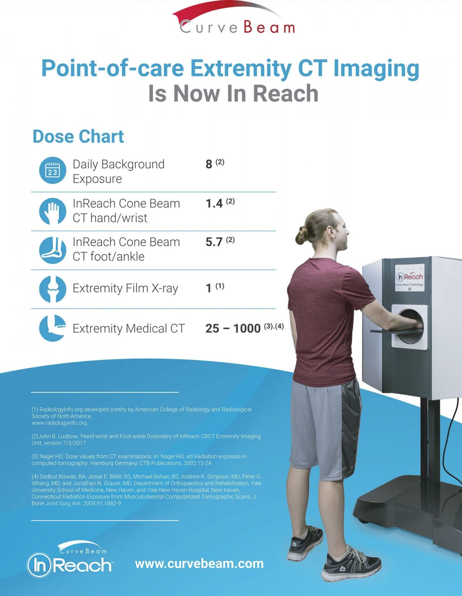
Radiation has a bad reputation, especially among lay people, but even in the medical community to a degree. Radiation is in the air we breathe, the food we eat, the water we drink, and even in our own inner biological…