Independent Research Organizations

The WBCT Society promotes dialogue and collaboration on WBCT research initiatives, and is working to create standardized protocols for WBCT measurements and analysis. Click here to learn more.
CT in Osteoarthritis Research (OCTA) has a goal of developing standard CT imaging protocols for osteoarthritis research. Click here to learn more.
Other Resources
Articles & White Papers
.
Popular Articles

Cone beam CT: A technical explanation of white paper image quality characteristics
Synopsis: Cone Beam Computed Tomography (CBCT) Advanced Imaging Systems are designed to capture three-dimensional weight bearing and non-weight bearing volumetric images of the body extremities. CBCT images are initially acquired as two-dimensional projections, using a rotating gantry with a relatively low power X-Ray source, a pulsed X-Ray beam, and a flat panel detector

Can Weight Bearing CT Advance our Planning for Forefoot Reconstructive Surgery?
Synopsis: The fact that the foot and ankle can withstand such high pressures and weight levels without failure over the course of a lifetime is truly amazing. The foot and ankle have such diverse shapes, sizes and mechanical positions in different people, but still tend to function at such a high level. It would be interesting to consider the advancements in architecture and compare them to the potential advancements in foot and ankle surgery, and how the thinking of both should be similar.

The 3D Advantage: Weight Bearing CT vs. X-Ray
Synopsis: CurveBeam AI Cone Beam CT (CBCT) systems are designed to capture three-dimensional weight bearing and non-weight bearing volumetric images of the body extremities. Cone Beam CT images are initially acquired as two-dimensional projections, using a rotating gantry with a fixed-anode X-Ray tube ring, a pulsed X-Ray beam, and a flat panel detector. The gantry rotates 360 degrees and acquires image projections, which are then reconstructed to create a series of axial slices.
More Articles
Excerpt: Podiatric physicians and surgeons routinely evaluate the osseous structures in their scope when performing diagnosis and treatment. After careful clinical examination where multiple diagnoses are considered, the differential list often leans to radiography for narrowing the diagnosis or confirmation of a diagnosis. Confirmation of osseous healing, whether post-injury or post-operatively (post osteotomy, arthrodesis, etc.), requires visualization of the bone. Likewise, in the study of lower extremity deformities, radiography is indispensable for comprehensive management. Aside from injury and disease states, very often baseline radiographs may be ordered on neuropathic patients, athletes, or in pediatric patients for growth evaluation. These are all reasons why most physicians who routinely treat foot and ankle conditions have in-office X-ray machines.
Excerpt: Evaluation of the condition we commonly call a ‘bunion’ is a rather complex process most podiatric physicians perform routinely. Many patients will present to the clinic with complaints of pain or pressure from “the bump on the inside of the foot.” Of course, they will often call it a bump., bunion, or spur, but I have not yet had any patients present themselves complaining of their “hallux abducto-valgus deformity.” Yet immediately, as a foot and ankle surgeon, I translate the “bump” into a triplane orthopedic deformity having osseous and soft tissue components and involving four separate bones. Interesting how quickly that happens when you think about it.
Excerpt: When evaluating the patient with metatarsalgia there are many anatomic and biomechanical factors to consider: first ray hypermobility, metatarsal length, metatarsal elevation and metatarsophalangeal joint stability (plantar plate). Traditional weight bearing radiographs can evaluate metatarsal length, but cannot assess metatarsal sagittal plane position. Sesamoid axial views can be used to evaluate the metatarsal head sagittal plane position, but such radiographs are not reliable as the patient or the x-ray technician positions the foot that clearly does not represent a resting stance position. Weight bearing CT can accurately measure the relative position of the metatarsal heads to one another and to the weight bearing surface.
Excerpt: Osteomyelitis is one of the most feared complications of diabetic foot ulceration, which often leads to lower extremity amputation and disability. Early diagnosis of osteomyelitis increases the likelihood of successful treatment and allows to minimize surgical resection and preserve ambulatory function. Unfortunately, most of the currently available imaging modalities are of limited use in assessing early stages of bone infection due to their low specificity and sensitivity for early osteolytic changes. Magnetic resonance imaging, though sensitive and specific, is very costly, which precludes its use during initial assessment of diabetic foot infection. In this report, we describe the use of a novel portable cone-beam computed tomography (CBCT) device, PedCAT, which may serve as a relatively inexpensive, accurate and readily available tool for early diagnosis of osteomyelitis in a diabetic patient. The success of this imaging modality is illustrated by two case studies, in which the use of PedCAT allowed us to diagnose and treat osteomyelitis in a timely manner, before any signs of bone destruction became apparent on plain radiographs. Though this report focuses on osteomyelitis, we believe that portable CBCT scanners hold unlimited potential in facilitating an accurate diagnosis of many musculoskeletal deformities of lower extremities, and would make a useful addition to an arsenal of imaging modalities currently available to a podiatric surgeon.
Excerpt: Avascular necrosis of the second metatarsal head can be a very difficult and troubling issue. The problem often occurs in children or teenaged athletes, and can make the continuation of sports activity difficult or impossible at times. We do not fully understand the underlying cause of the avascular changes but often consider chronic microtrauma to be a common source. A long metatarsal may also contribute to the underlying pressure and eventual cartilage collapse. If we pick it up early, the problem is commonly easier to treat. With time, there is collapse of the articular surface, making treatment more difficult.
Excerpt: What is three-dimensional cone beam imaging? Unlike conventional computed tomography (CT), which has a fan-shaped X-ray beam, modalities such as the pedCAT (Curvebeam) have a cone-shaped X-ray beam. This allows clinicians to obtain an image of a volume of tissue in one circumferential pass instead of having to take multiple slices with multiple exposures.
Excerpt: I now routinely take a WBCT scan six weeks after joint fusion surgery to measure the exact degree of healing. I now also CT every ankle fracture, and I have been surprised at the variability. Especially posterior fractures – they are often bigger than they appeared on plain X-Ray, and they often travel around to the medial side as well, which we never knew they did. For years, we had no reproducible, reliable measurement of hindfoot alignment. Now you can scroll through the tibia, talus and calcaneus through different planes and draw your alignment measurement easily. I tell my colleagues all the time, it’s hard for me now to order a plain X-Ray because I feel I’m not getting enough information.”
Download case study here
Download case study here
Download case study here
Download case study here
Download case study here
Download case study here
Download case study here
Download case study here
Popular Weight Bearing CT Studies

Synopsis: The use of preoperative and intraoperative guidance in foot and ankle surgery has grown substantially in recent years. Weight-bearing computed tomography (WBCT) and patient-specific instrumentation (PSI) are used in total ankle arthroplasty (TAA) to achieve precise bone cutting and implant positioning, and intraoperative 3-dimensional (3D) imaging has been used to reduce complications and improve clinical outcomes in other foot and ankle surgical procedures. This narrative review of the literature focuses on the evidence supporting the use of WBCT and PSI in TAA and looks at other promising technologies used to guide foot and ankle surgery.

Weight Bearing Cone Beam CT Scans and It’s Uses in Ankle, Foot, and Knee: An Update Article
Synopsis: Imaging plays a key role in the preoperative diagnosis, surgical planning, and postsurgical assessment of the foot, ankle, and knee pathologies. Interpreting diagnostic imaging accurately is crucial for the clinical practice of orthopedic surgeons. Among the most used imaging modalities radiographic assessments are amenable to errors for various technical reasons and superposition of bones. CT is a conventional imaging procedure that provides high-res images, but fails in considering a truly WB condition. In an attempt to overcome this limitation, WB CBCT technology has being successfully employed in the clinical practice for the past decade. Besides economically viable and safe, the WB cone beam CT considers WB conditions and provides high-quality scans.

Weight-bearing CT in foot and ankle pathology
Synopsis: Cone-beam scanners (CBCT) enable CT to be performed under weight-bearing – notably for the foot and ankle. The technology is not new: it has been used since 1996 in dental surgery, where it has come to replace panoramic X-ray. What is new is placing the scanner on the ground, so as to have 3D weight-bearing images, initially of the foot and ankle, and later for the knee and pelvis. This saves time, radiation and money. It is now increasingly used, but is unfortunately limited by not having specific national health insurance cover in France, and by the psychological reticence that goes with any technological breakthrough. A review of the topic is indispensable, as it is essential to become properly acquainted with this technique. To this end, we shall be addressing 5 questions. What biases does conventional radiography incur?
More Weight Bearing CT Studies
Excerpt: Musculoskeletal radiology has been mostly limited by the option between imaging under load but in two dimensions (i.e., radiographs) and three-dimensional (3D) scans but in unloaded conditions (i.e., computed tomography [CT] and magnetic resonance imaging in a supine position). Cone-beam technology is now also a way to image the extremities with 3D and weight-bearing CT. This article discusses the initial experience over a few studies in progress at an orthopaedic center. The custom design of total ankle replacements, the patellofemoral alignment after medial ligament reconstruction, the overall architecture of the foot bones in the diabetic foot, and the radiographic assessment of the rearfoot after subtalar fusion for correction of severe flat foot have all taken advantage of the 3D and weight-bearing feature of relevant CT scans. To further support these novel assessments, techniques have been developed to obtain 3D models of the bones from the scans and to merge these with state-of-the-art gait analyses.
Excerpt: Newer cone-beam computed tomography (CT) technology has grown in popularity for evaluation of foot and ankle pathology in the weight-bearing (WB) position. Many studies have demonstrated its benefits within the adult population, but there is a paucity of its use within the pediatric literature. The purpose of this study was to describe the indications and clinical findings of WBCT within a pediatric population.
Excerpt: Although 2D and 3D geometrical analyses are fundamentally different, automated 3D analyses are more reproducible and precise compared to 2D analyses. In addition, 3D evaluation better demonstrates differences in bone configurations between weight-bearing and non weight-bearing conditions, which may be of value to demonstrate pathology.
Consensus for the Use of Weightbearing CT in the Assessment of Progressive Collapsing Foot Deformity
Excerpt: There is evidence that the use of WEIGHTBEARING imaging aids in the assessment of progressive collapsing foot deformity (PCFD). The following WEIGHTBEARING conventional radiographs (CRs) are necessary in the assessment of PCFD patients: anteroposterior (AP) foot, AP or mortise ankle, and lateral foot. If available, a hindfoot alignment view is strongly recommended. If available, WEIGHTBEARING computed tomography (CT) is strongly recommended for surgical planning. When WEIGHTBEARING CT is obtained, important findings to be assessed are sinus tarsi impingement, subfibular impingement, increased valgus inclination of the posterior facet of the subtalar joint, and subluxation of the subtalar joint at the posterior and/or middle facet.
Excerpt: Weight-bearing makes greater the three-dimensional deformities in knees with osteoarthritis. Particularly, greater tibial internal rotation was observed in patients with grades 2 and 3 compared to those with grade 1. The greater tibial internal rotation due to weight-bearing is a key pathologic feature to detect early osteoarthritic change in knees undergoing osteoarthritis.
Excerpt: Total ankle arthroplasty (TAA) is a popular and viable option for end-stage ankle arthritis. Posttraumatic arthritis is the most common etiology of ankle arthritis, which creates the additional challenge of osseus deformity. Accuracy and reproducibility in placing the implant on the mechanical axis has been shown to be paramount in all joint arthroplasty including total ankle replacement. Patient-specific preoperative navigation is a relatively new technology for TAA, and up until this past year has been based off of nonweightbearing (NWBCT) or simulated weightbearing computed tomography (WBCT). Our institution has created a protocol to use WBCT in the preoperative patient-specific navigation for TAA using the Prophecy system. The purpose of our study was to compare the accuracy and reproducibility of implant alignment and size using WBCT vs prior studies using NWBCT for the Prophecy reports.
Excerpt: Weight-bearing CT (WBCT) scans of the foot and ankle have improved the understanding of deformities that are not easily identified on radiographs and are increasingly being used by orthopaedic surgeons for diagnostic and preoperative planning purposes. In contrast to standard CT scans, WBCT scans better demonstrate the true orientation of the bones and joints during loading. They have been especially useful in investigating the alignment of complex pathologies such as adult-acquired flatfoot deformity in which patients have been found to have a more valgus subtalar joint alignment than in a normal cohort and high rates of subfibular impingement. Studies using WBCT scans have also provided new insight into more common lower extremity conditions such as hallux valgus, ankle fractures, and lateral ankle instability.
Excerpt: We investigated the association between hindfoot residual malalignment assessed on weightbearing computed tomography (WBCT) images and the development of periprosthetic cysts (PPCs) after total ankle replacement (TAR). We hypothesized that PPCs would be found predominantly medially in the varus configuration and laterally in the valgus configuration.
1
Popular Cone Beam CT Studies

Synopsis: An analysis of the capabilities of cone-beam computed tomography (CBCT) in the assessment of the form and structure of wrist and hand bones was the aim of the research. CBCT of wrist and hand was conducted in a group of voluntary patients, which included 40 members aged 22- 68 years. According to the results of the research, CBCT shows a high efficiency in detection of form, measurements and structural changes of bones of the anatomic region.

Cone-beam CT for Diagnosis of Avulsion Injuries of the Hand and Wrist
Synopsis: Unlike multidetector CT, where a fan-shaped beam moves helically around the patient and falls on a linear detector, cone-beam CT uses a cone- or pyramid-shaped beam projected on a flat panel detector. This design allows the entire volume to be imaged in a single rotation with lower radiation dose than that of multidetector CT and a high spatial resolution (as low as 75 µm), with a trade-off of reduced soft-tissue contrast resolution and greater risks of motion artifact (2).

Synopsis: During 2015, a new model of CBCT, New Tom 5G XL (Verona), was used in the Radiology Department of The University of Verona, in collaboration with the Orthopaedics Department. The study compared CBCT with standard x-ray in the diagnosis of foot and ankle fractures, tibial plateau fractures, wrist and scaphoid fractures, elbow fractures. We can say that if compared to standard X-ray, CBCT has higher sensitivity and specificity in the proper identification and typing of these kind of lesions, with low exposition dose if compared to MDCT.
More Cone Beam CT Studies
Excerpt: Incorporating cone-beam CT into routine clinical practice as part of a standardized diagnostic algorithm yielded a 50% fracture detection rate in patients with negative wrist radiographs but ongoing clinical concern for radiocarpal fracture. Cone-beam CT provides more diagnostic information than radiographs at a lower radiation dose than conventional MDCT. Given the poor accuracy of radiographs for acute radiocarpal fractures and the high fracture prevalence in this cohort, we feel that cone-beam CT should be regarded as the new standard of care in the investigation of these patients.
Excerpt: The objective was to compare the diagnostic accuracy and radiation exposure of cone beam computed tomography (CBCT) and multislice computed tomography (MSCT) in the evaluation of finger fractures. The results found cone beam CT may be considered a valuable imaging tool in the preoperative assessment of finger fractures, when MSCT is not available.
Excerpt: Scaphoid fractures are challenging to accurately diagnose with delayed and missed diagnoses risking poor patient outcomes. Cone beam CT (CBCT) is an emerging technology facilitating alternative access to multi-planar imaging. The aim of this study was to evaluate the use of early CBCT in the diagnosis of suspected scaphoid fractures presenting via the Emergency Department (ED). CBCT heralds the potential for early accurate diagnosis of radiocarpal fractures, at lower cost, shortening clinical pathways and reducing clinical risk in the ED.
Excerpt: The objective is to evaluate the diagnostic value of cone beam computed tomography (CBCT) for scaphoid and wrist fractures that are missed on standard radiographs. The results demonstrated CBCT is superior to radiographs for diagnosing occult cortical fractures. Because of its low radiation dose, we believe that CBCT can be used in current practice as a replacement or supplement to radiographs to detect these fractures and optimize the cost-effectiveness ratio by limiting the number of needless immobilizations.
Excerpt: Weight-bearing makes greater the three-dimensional deformities in knees with osteoarthritis. Particularly, greater tibial internal rotation was observed in patients with grades 2 and 3 compared to those with grade 1. The greater tibial internal rotation due to weight-bearing is a key pathologic feature to detect early osteoarthritic change in knees undergoing osteoarthritis.
1
Popular Financial / Operational Studies and White Papers

Cost Analysis and Utilization of Weight Bearing CT
Synopsis: Weightbearing Computed Tomography (WB CT) is becoming a valuable tool in the evaluation and understanding of foot and ankle pathology. With this, more practices may be interested in acquiring WB CT scanners and understanding cost effectiveness of acquisition. To date there are currently no US based reports of cost analysis of WB CT scanners. Our study is a cost analysis of WB CT at tertiary referral physician owned practice.

MGH Study Finds WBCT Best Option for Patients
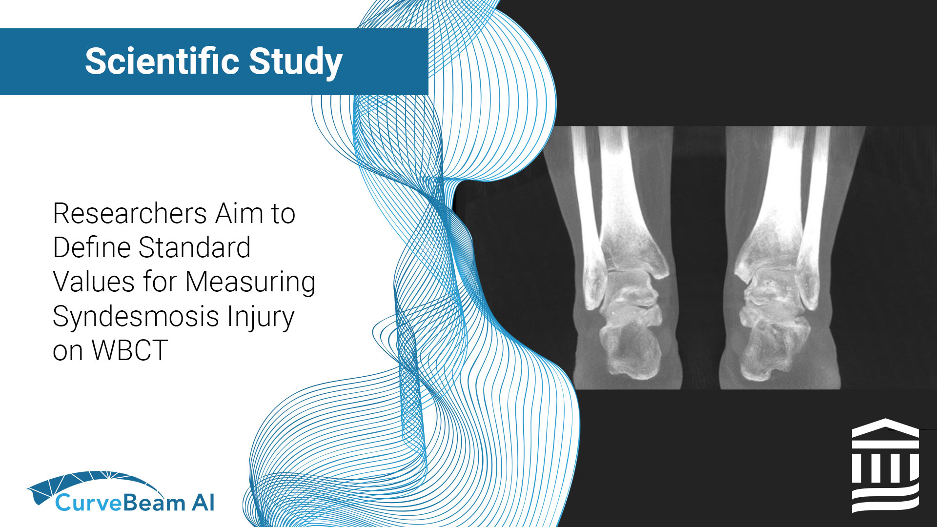
Synopsis: Using a weight bearing CT (WBCT)-first approach for diagnosing syndesmotic instability is the most cost-effective option for patients, researchers at the Foot and Ankle Research Innovation Lab (FARIL) at Massachusetts General Hospital found in a recent study. FARIL researchers presented these findings via a poster at the recent 2021 AOFAS Annual Meeting in Charlotte, N.C. Accurate and timely diagnosis of syndesmotic instability is critical for optimal clinical outcomes, but subtle instability can be challenging to identify. WBCT has proven to be a reliable tool for diagnosing syndesmotic instability under physiologic load.

Synopsis: WBCT has been proven to more precisely measure bone position than conventional weightbearing radiographic series (R) and conventional CT (CT). The purpose of this study was to assess the benefit of using WBCT instead of R and/or CT as the standard imaging modality, evaluating image acquisition time, radiation dose, and cost-effectiveness. The study resulted in 10% decreased RD, 77% decreased T, and increased financial profit (51 Euros per patient) for the institution.
More Financial/ Operational Studies and White Papers
Excerpt: The purpose of this study was to evaluate the cost-effectiveness of introducing cone-beam computed tomography (CBCT) in the management of the complex finger fractures with articular involvement. We created a decision tree model simulating the diagnostic pathway of complex finger fractures, suggesting the use of CBCT as alternative to multi-slice computed tomography (MSCT), and we compared their clinical outcomes, costs, and cost-effectiveness for a hypothetical cohort of 10,000 patients. Measures of effectiveness are analysed by using quality-adjusted life years, incremental cost-effectiveness ratio, and net monetary benefit.
Bone Health Studies
.
Popular Studies

Synopsis: More than 70% of women sustaining fractures have osteopenia or “normal” bone mineral density (BMD). As microstructural deterioration increases bone fragility disproportionate to the bone loss producing osteopenia/normal BMD, we hypothesized that the SFS of ≥70 units, a measure capturing severe cortical and trabecular deterioration, will identify these women.

Synopsis: The disease of bone fragility is captured by cortical and trabecular deterioration: A measurement of coexisting cortical and trabecular deterioration is likely to identify women at risk for fracture more robustly than absolute values of cortical porosity, trabecular density, or BMD.
 2021")
Synopsis: Treatment is usually withheld from women with osteopenia even though they are the source of over 70% of all women having fragility fractures. As microstructural deterioration increases fracture risk and zoledronate reduces it, we aimed to determine whether identifying and treating women with osteopenia and severe microstructural deterioration is cost-effective.
More Studies
Excerpt: Investigation identified an elevated serum tryptase, and marrow biopsy confirmed the diagnosis of mastocytosis. Lactation causes bone loss, but the occurrence of fractures in the setting of severe deficits in BMD and microstructural deterioration signals the need to consider additional causes of bone loss.
Excerpt: After menopause, remodeling becomes unbalanced and rapid. Each of the many remodeling transactions deposits less bone than it resorbed, producing microstructural deterioration. Trabecular bone is said to be lost more rapidly than cortical bone. However, because 80% of the skeleton is cortical, we hypothesized that most menopause-related bone loss and changes in bone microstructure are cortical, not trabecular in origin, and are the result of intracortical remodeling. Distal tibial and distal radial microstructure were quantified during 3.1 years (range, 1.5 to 4.5 years) of follow-up using high-resolution peripheral quantitative computed tomography and StrAx software in 199 monozygotic and 125 dizygotic twin pairs aged 25 to 75 years in Melbourne, Australia.
Excerpt: Reduced bone mineral density (BMD) may be due to reduced mineralized bone matrix volume, incomplete secondary mineralization, or reduced primary mineralization. Because bone biopsy is invasive, we hypothesized that noninvasive image acquisition at high resolution can accurately quantify matrix mineral density (MMD). Quantification of MMD was confined to voxels attenuation photons above 80% of that produced by fully mineralized bone matrix because attenuation at this level is due to variation in mineralization, not porosity. Low-radiation HR-pQCT may facilitate noninvasive quantification of bone’s MMD and microstructure in health, disease, and during treatment.
Excerpt: The severe cortical porosity and trabecular deterioration associated with estradiol depletion and the longevity of premenopausal women with early breast cancer treated with endocrine therapy provide a compelling rationale to investigate the efficacy of antiresorptive therapy initiated at the time of breast cancer treatment.
Excerpt: Assessment of sex, race and age related differences in microstructure requires measurement of anatomically equivalent regions.
Excerpt: Effects of intermittent teriparatide depend on the dose and frequency of administration. Daily dosing, particularly the higher dose, but not weekly dosing, increased cortical porosity. Work is needed to investigate the effects of the regimens on bone formation.
Excerpt: Bone microstructure may be irreversibly deteriorated after cessation of breastfeeding at appendicular sites. Studies are needed to establish whether this deterioration compromises bone strength and increases fracture risk later in life.
Excerpt: Denosumab reduced cortical porosity of the proximal femoral shaft, resulting in increased mineralized matrix volume and improved strength, changes that may contribute to the reduction in hip and nonvertebral fractures reported with denosumab therapy.
Excerpt: We report that a postmenopausal woman with osteoporosis developed bilateral incomplete atypical femoral fractures (AFFs) after seven years of bisphosphonate therapy. Cessation of the bisphosphonate and treatment with teriparatide was associated with near complete radiological resolution of the AFFs. After 12 months without treatment, denosumab was commenced to prevent structural deterioration. Six months later she developed recurrent bilateral AFFs. This case highlights the management dilemma in patients with ongoing bone loss but prone to stress fractures associated with antiresorptive therapy. Stopping the antiresorptive is recommended but structural decay will recur predisposing to fragility fractures. If the antiresorptive is continued, bone material composition will be further compromised predisposing to atypical fractures. Teriparatide may assist healing of stress fractures and improvement in bone matrix composition. Later antiresosrptive therapy to preserve bone microstructure may compromise material composition.
Excerpt: Increased BTM levels are associated with higher cortical porosity, thinner cortices, larger bone size and higher odds for fracture. We infer that this is produced by increased periosteal apposition, intracortical and endocortical remodeling; and that these changes in bone architecture are predisposing to fracture.
Excerpt: In women with osteoporosis, porosity is captured by aBMD and so measuring UDR cortical porosity does not improve diagnostic sensitivity. However, in women with osteopenia, cortical porosity was associated with forearm fractures.
Excerpt: These data suggest that structural decay owing to bone remodeling and progression of bone fragility may be prevented more effectively with denosumab.
Excerpt: Between-group differences were significant for compact-appearing cortex porosity (p = 0.005). Risedronate slows microstructural deterioration in younger and partly reverses it in older postmenopausal women, features likely to contribute to antifracture efficacy.
Excerpt: Taller women are at increased risk for fracture despite having wider bones that better tolerate bending. Because wider bones require less material to achieve a given bending strength, we hypothesized that taller women assemble bones with relatively thinner and more porous cortices because excavation of a larger medullary canal may be accompanied by excavation of more intracortical canals. Three-dimensional images of distal tibia, fibula, and radius were obtained in vivo using high-resolution peripheral quantitative computed tomography (HRpQCT) in a twin study of 345 females aged 40 to 61 years, 93 with at least one fracture.
Excerpt: Segmentation of the transitional zone minimizes errors in apportioning cortical fragments and cortical porosity to the medullary compartment and so is likely to allow accurate assessment of fracture risk and the morphological effects of growth, aging, diseases and therapies.
Excerpt: Osteoporosis research has focused on vertebral fractures and trabecular bone loss. However, non-vertebral fractures at predominantly cortical sites account for 80% of all fractures and most fracture-related morbidity and mortality in old age. We aimed to re-examine cortical bone as a source of bone loss in the appendicular skeleton.
TEXTBOOKS
.

Weight Bearing Cone Beam Computed Tomography (WBCT) in the Foot and Ankle – A Scientific, Technical and Clinical Guide, edited by Martinus Richter, Francois Lintz, Cesar de Cesar Netto, Alexej Barg, Arne Burssens, Scott Ellis. Published by Springer Nature

3-D orthopaedic pre-operative surgical planning for total ankle replacement”, appearing in, Primary and Revision Total Ankle Replacement: Evidence-Based Surgical Management, Second Edition, edited by, Thomas S. Roukis, Gregory C. Berlet, Christopher Bibbo, Christopher F. Hyer, & Murray J. Penner. Published by Springer Nature

McGlamry’s Comprehensive Foot and Ankle Surgery, 5th edition, edited by, Carpenter Published April 2021, by Walters Kluwer

Controversies in Managing the Progressive Collapsing Foot Deformity (PCFD), An issue of Foot and Ankle Clinics of North America, Volume 26-3, edited by Cesar de Cesar Netto. Published by Elsevier

Le grandi deformità della caviglia e del piede Vol. 23 SICP, Authors: Federico Giuseppe Usuelli, Francesco Ceccarelli, Sandro Giannini, Umberto Alfieri Montrasio
News & Events

AI-Driven Segmentation on Weight Bearing CT Introduction Accurate segmentation is critical for orthopedic workflows, including…

Orthopedic decision-making depends on accurate representation of anatomy—particularly when joint alignment and bone relationships change…

Fragility fractures are often the first visible sign of underlying osteoporosis but too often, they…
In recent years, patient-specific instrumentation (PSI) for total ankle arthroplasty (TAA) has gained momentum, driven…

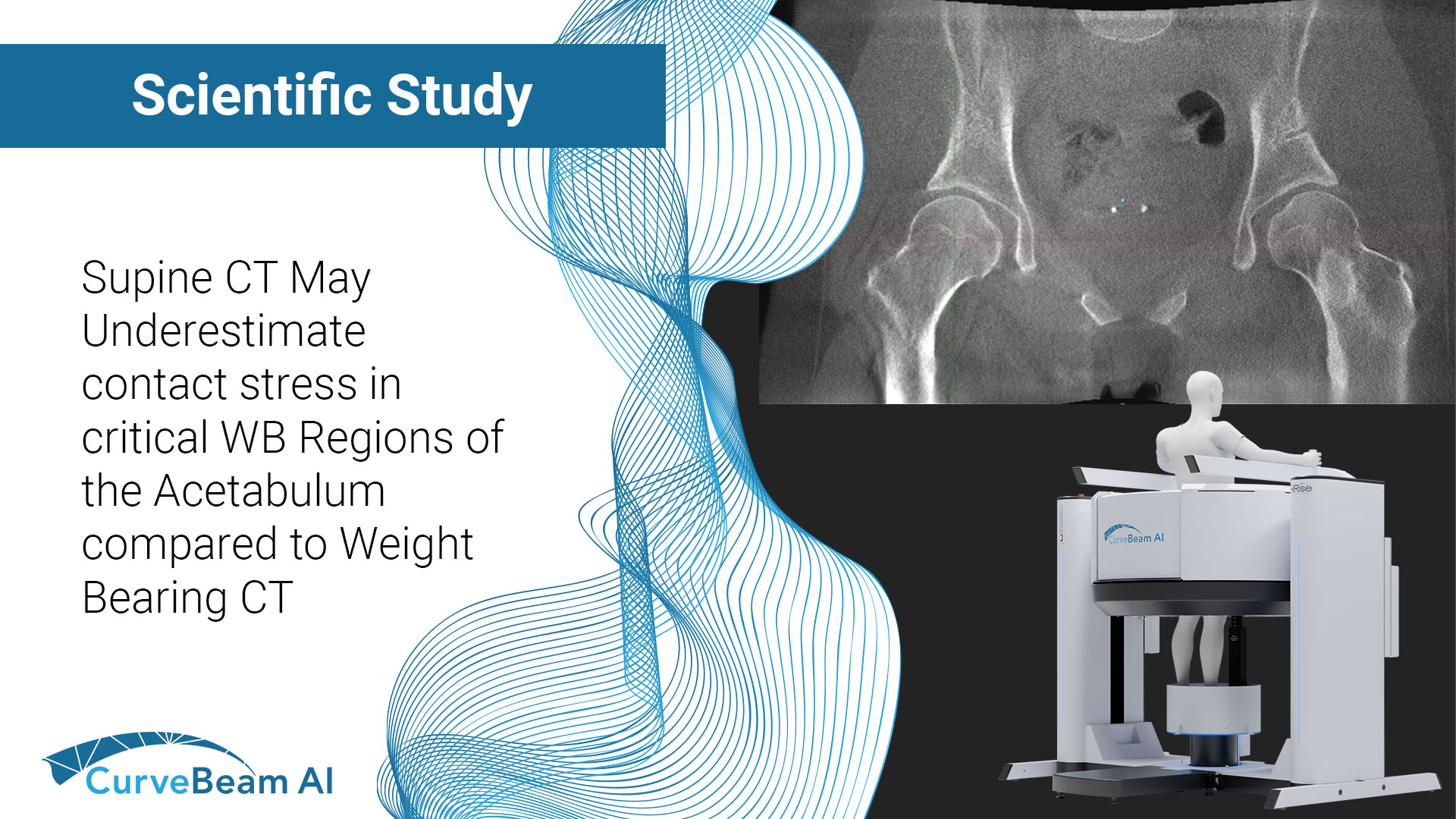
Key Points: Computational models of the hip often omit patient-specific functional orientation when placing imaging-derived…
Key Points: Weight Bearing CT (WBCT) allows for bilateral comparative data that is imperative when…
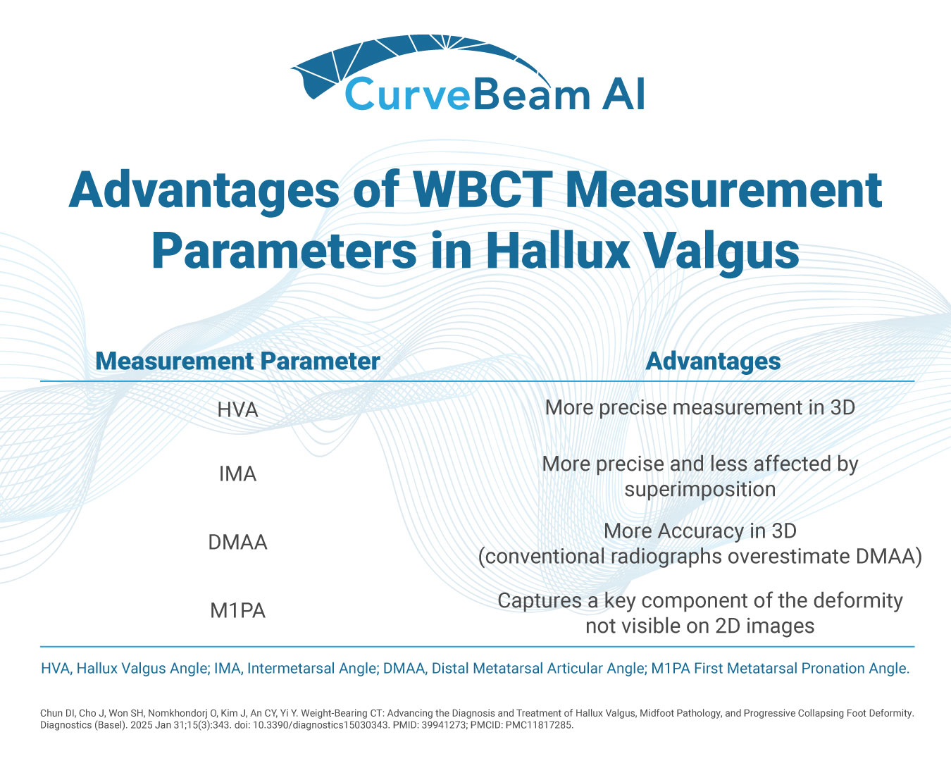
When it comes to hallux valgus (bunion) deformity, weight bearing CT provides significant advantages over…
