AI-Driven Segmentation on Weight Bearing CT Introduction Accurate segmentation is critical for orthopedic workflows, including…
Implementing Automated 3D Measurements to Quantify Reference Values and Side-to-Side Differences in the Ankle Syndesmosis
Key Points:
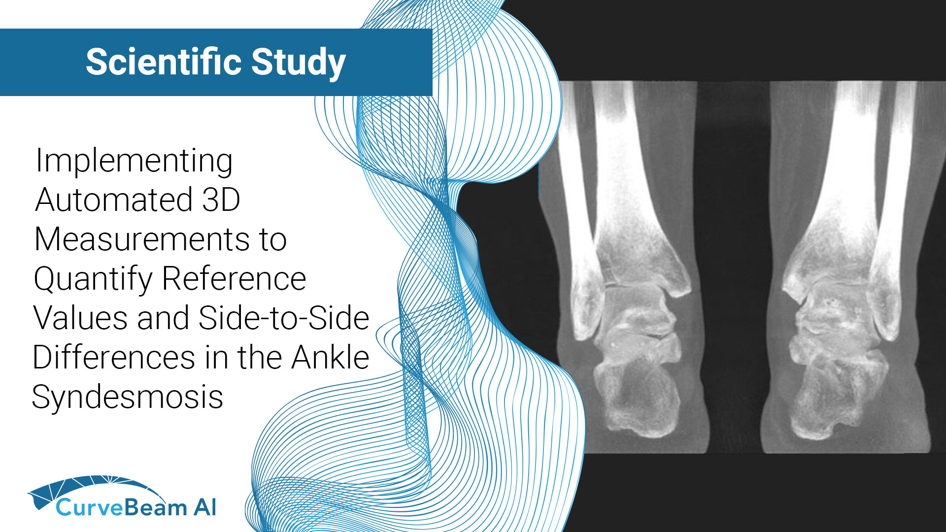
- Currently plain radiographs are the standard method in diagnosing syndesmotic ankle injuries even though the distal tibiofibular joint cannot be assessed due to superposition of the osseous structures in the foot.
- Weight bearing CT (WBCT) allows bilateral assessment of the ankle syndesmosis while under physiological load and in 3-dimensions (3D).
- Researchers created a novel algorithm that is able to utilize WBCT imaging to define normative reference values, allowing them to capture alignment of the ankle syndesmosis in all six degrees of freedom which vastly improves upon current 2-dimensional (2D) methods.
Syndesmotic ankle injuries are present in up to 18% of all ankle sprains and up to 20% of ankle fractures requiring internal fixation. When left undiagnosed and/or untreated, they may lead to syndesmotic instability and subsequently post-traumatic ankle osteoarthritis, which can be challenging to diagnose, especially when subtle.
Current diagnostics utilize plain radiographs and manual measurements, comparing the injured distal tibiofibular joint to the non-injured side. Current literature is unclear on whether the configuration of the ankle syndesmosis is symmetrical and certain types of displacement such as rotation within the distal tibiofibular joint cannot be assessed on plain radiography due to superposition of the osseous structures. Current techniques are also cumbersome to employ and often not integrated into the viewing programs commonly used in today’s clinical practices. While Conventional CT (MDCT) imaging overcomes these issues and allows different angular measurements to quantify rotation, weight bearing CT improves upon MDCT even further by allowing a bilateral assessment of the ankle syndesmosis while under physiological load in 3-dimensions.
Dr. Peiffer et. al out of the Harvard Medical School, Massachusetts General Hospital, Boston, MA, USA, conducted a study with the objective to determine physiological reference values concerning the 3D configuration of the normal ankle syndesmosis based on an automated method (measurements that are not yet established), as well as determine whether the ankle syndesmosis is symmetrical across all 3D measurements on both weight bearing and non-weight bearing (NWBCT) imaging. They found that by utilizing a novel algorithm, which was trained to automate the segmentation step using artificial intelligence (AI) along with manual measurements between the non-injured and injured sides symmetry, that in a clinical setting this new algorithm could surmount the current limitations of manual 2D measurements and distinguish patients with a syndesmotic ankle lesion from normal variance.
Researchers used data from 76 NWBCT ankles (retroactive study) and 86 WBCT ankles. WBCT scans were obtained using CurveBeam AI’s pedCAT system. Results showed no statistically significant differences could be detected between 3D measurements of left and right ankles for all variables.
It was concluded that their novel automated 3D algorithm was able to define normative references values and revealed a side-to-side symmetry which allowed them to capture the alignment of the ankle syndesmosis in all six degrees of freedom, improving upon previously established measurements on 2D plain radiographs or CT image slices.
The authors noted that future research could improve the present study by using prospectively recruited cohorts with additional clinical and MRI investigations to assure an intact condition of the syndesmotic ligaments. It could also be assessed if their 3D techniques could aid in the treatment of syndesmotic ankle injuries by correcting the amount of calculated deviation relative to a patients uninjured side, e.g. in case of a malrotated or shortened fibula.
To read the full study click here.
Related Posts


