Read the article.

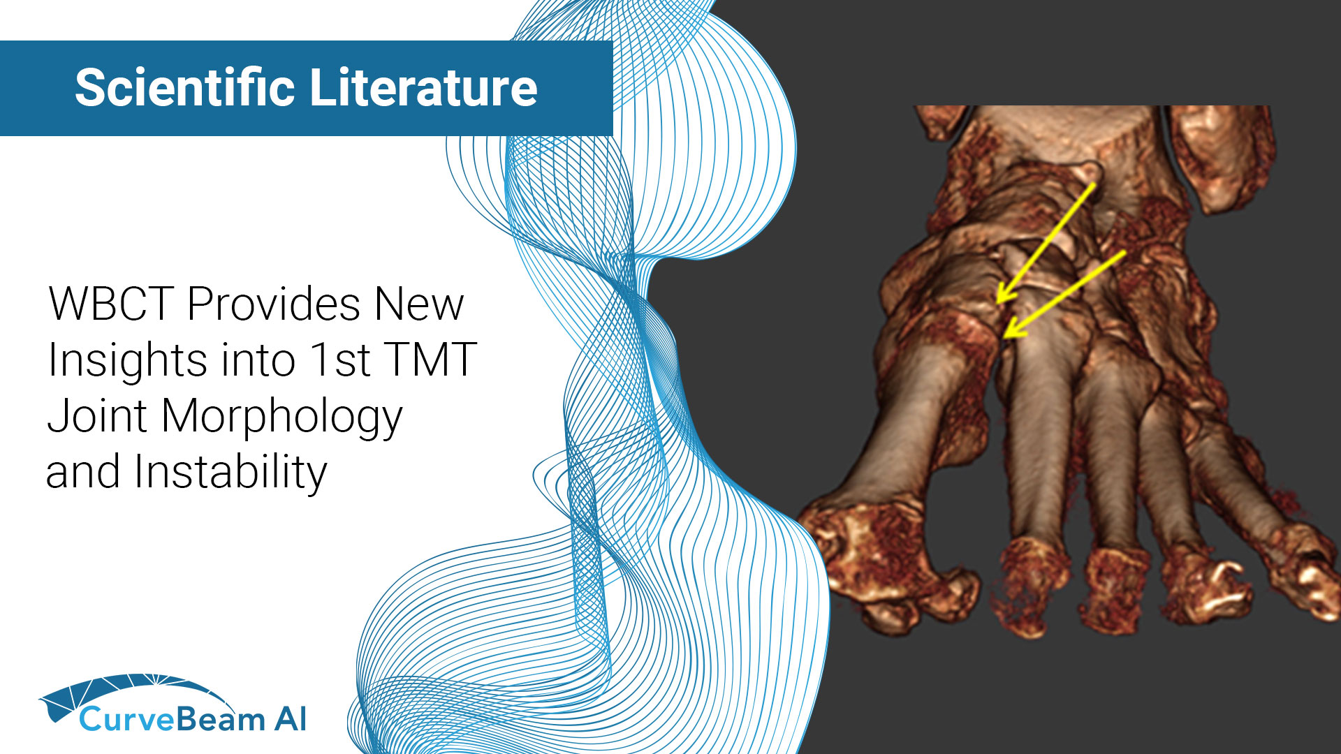
Case-Control Study: The Role of First Tarsometatarsal Joint Morphology and Instability in the Etiology of Hallux Valgus
Key Points:
- First tarsometatarsal joint (TMT1) hypermobility is associated with hallux valgus (HV) which is caused by TMT1 instability.
- Weight Bearing CT (WBCT) provides a clearer view of TMT1 morphology and instability by providing images of a patient under physiological load.
The TMT1 is a crucial component of the medial column of the foot and plays a significant role in first-ray mechanics. Researchers found that first-ray hypermobility primarily arises from TMT1 instability, though there is still a limited understanding of TMT1 morphology and anatomy due to the challenges and limitations of radiographic imaging.
Dr. Linfeng, MD et al from the Department of Foot and Ankle Surgery, Beijing Tongren Hospital at the Capital Medical University in Beijing, China aimed to investigate the TMT1 morphology and its potential correlation with TMT1 instability and the occurrence of HV using WBCT. WBCT allows for the assessment of the foot under physiologic load and enables measurement of 3D kinematics of TMT1 morphology.
Case Report
82 consecutive feet with HV that underwent WBCT scans, as well as 70 controls, were reviewed in this case-control study. Researchers found that the middle facet width (MFW), lower inferior lateral facet angle (ILFA), and inferior lateral facet height (IFLH) in TMT1 were associated with more severe HV. Additionally, the continuous-flat type of TMT1 mostly presented in HV patients, whereas the separated-protruded type primarily appeared in the controls. A new possible association between the continuous-flat type of TMT1 and TMT1 instability was also found, providing new insights into TMT1 instability and HV in the aspect of TMT1 anatomy.
Researchers noted that further larger sample prospective studies are necessary to investigate the clinical outcome and postoperative recurrence rates of different TMT1 morphology types.
To read the full study click here.


