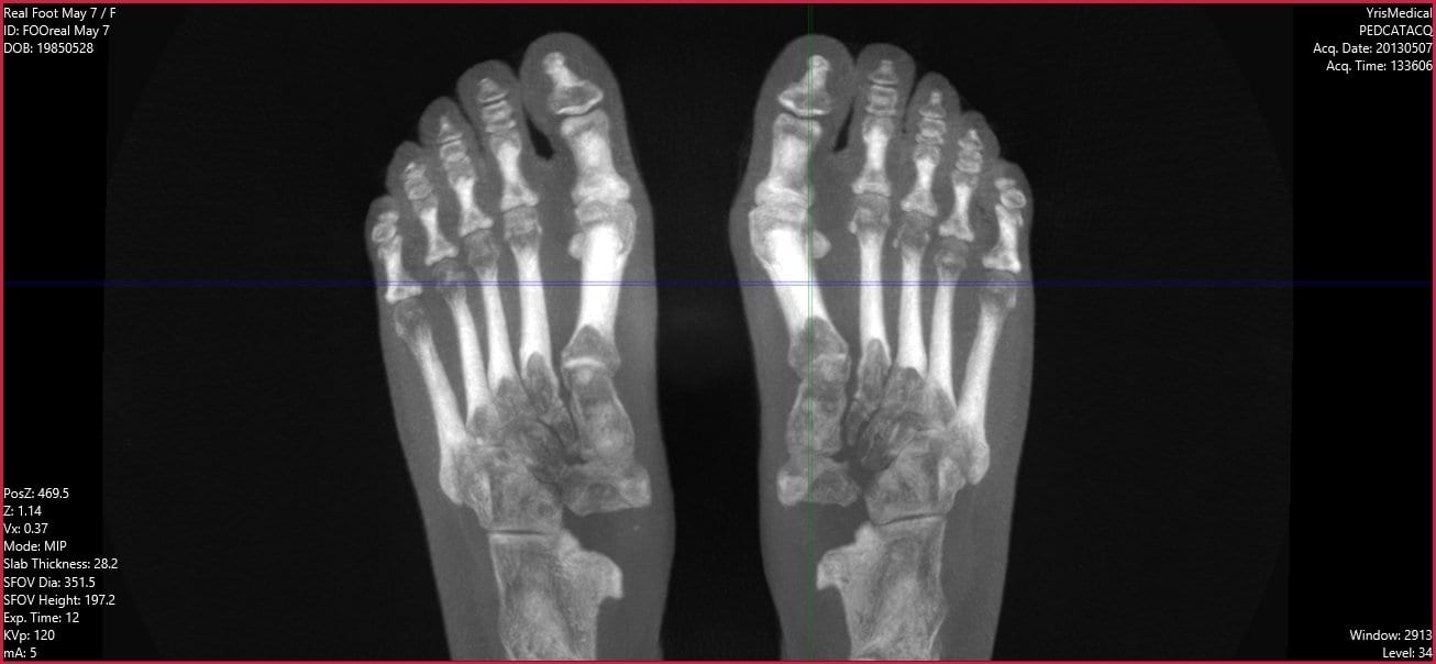
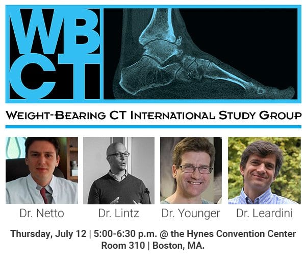
Chat with us, powered by LiveChat Skip to content

pedCAT: A Positive User Experience

“After using the pedCAT for a year, I am very impressed with how much I need it, and how many different things I use it for,” Dr. Bob Baravarian said at the 2015 Western Foot and Ankle Conference held in Anaheim, Cali. The California Podiatric Medical Association hosted the meeting at the end of June. Dr. Baravarian cited Hallux Rigidus as one example where pedCAT imaging has been helpful because he is able to properly assess the sesamoids. Dr. Baravarian confirmed his practice is realizing positive revenue streams as a result of the device.
Read more

Diagnostic Options for Freiberg’s Avascular Necrosis

The underlying causes of avascular necrosis of the second metatarsal head are not totally understood, but early diagnosis is essential. Delayed treatment can result in a collapse of the articular surface, making treatment more difficult. Dr. Bob Baravarian, DPM, explained his preferred methods for diagnosing the condition in the May 2015 issue of Podiatry Today. An X-Ray will show the overall contour and alignment of the metatarsal head. MRI is one option for a secondary study, but edema and swelling can limit visualization of the surrounding bone region, Dr. Baravarian explained. Weight bearing CT scans, meanwhile, clearly show the bones and joints and how they are aligned. Weight bearing CT is "our go-to imaging study," Dr. Baravarian said in the article. "With adequate diagnostic testing and proper patient and procedure selection, one can treat avascular necrosis of the metatarsal head with good to excellent outcomes," Dr. Baravarian said. Dr. Baravarian is the director of University Foot and Ankle Institute in Los Angeles. His practice offers weight bearing CT imaging services.
Read more
ALT TEXT

Weight Bearing CT in Everyday Practice

“In my opinion, this will be the standard of care in the next couple of years. We are switching from two-dimensional technology to three-dimensional technology and this is really the future of not just foot and ankle, but the future of medicine,” Dr. Alex Tievsky, DPM, said in a lecture he gave at the Graham International Implant Institute 8th Annual Symposium in Miami, on April 17. 2D is beginning to be phased out,” Tievsky said. “Now we’re beginning to see the problem from all angles and all planes, so this is super helpful from that respect.” Dr. Tievsky presented a number of cases where he benefited from access to weight bearing 3D technology in his office. Clinical Case #1 A 50-year-old female presented with bilateral flat feet for 20 years. She had heard about the HyProCure procedure, which corrects hindfoot misalignment through a minimally invasive procedure. She was eager to have the procedure done, no matter the cost. Dr. Tievsky took a pedCAT scan, and found she had a severe talar coalition. talar calc coalition coronal left “How many times do you catch a coalition on an X-Ray? It’s hard,” Dr. Tievsky said. “Sometimes you can see a halo sign, but it’s often missed. On the first visit, I was able to tell her, we either have to resect this coalition or we have to do a fusion. It’s impossible to get this level of information on an X-Ray.” Clinical Case #2 A 16-year old girl came in with first metatarsal head pain. She had already been to two other podiatrists in the last six months, and they had prescribed steroid injections. The pedCAT revealed a fracture on her fibular sesamoid that is extremely easy to miss on X-Ray. fractured sesamoid “Within her first 10 minutes in the office, we had a diagnosis,” Dr. Tievsky said. “We treated her appropriately. We immobilized her for eight weeks and gave her a bone stimulator. And she was pain free, three podiatrists later. She was happy, her mom was happy, and she never came back.” Clinical Case #3 A patient presented with a lateral plantar fasciitis, a talo-tarsal dislocation, back pain for five years, knee pain, and hip pain. A pedCAT revealed she had a tumor in her bone and it was eroding it. “There was no way we would have caught it on X-Ray,” Dr. Tievsky said. “We sent her out for oncology. It could have a malignant tumor, and we could have saved her life.” Cyst Coronal The scan is quick to take and you have a full work-up in about three minutes, Dr. Tievsky said as a closing statement. “This is a super important tool,” he said. “I’m kind of biased. I love this now. I can’t practice without it.”
Read more

Weight Bearing CT Scans for the Evaluation of Implant Arthroplasty Candidates

Weight bearing CT scans can be critical to a proper diagnosis, even for routine procedures. In the following case, for example, a patient’s X-Rays indicated that he would be a good candidate for a metatarsal head hemi-implant arthroplasty. However, when the patient sought a second opinion, a weight bearing CT (pedCAT) scan revealed the true condition of the metatarsal head, and the surgical plan was considerably altered as a result. Click on the blog post title to read the case and view images.
Read more

Dr. Gary Briskin: The pedCAT is the Future

"It's the future - embrace it. It's your future - control it." Dr. Gary Briskin delivered this motivating message at the Foot & Ankle Business Innovations conference in Chicago on Jan. 23. Dr. Briskin, of University Foot & Ankle Institute in Santa Monica, Cali, discussed how the pedCAT weight bearing CT system has improved his practice since his group acquired one last year. His main take-away points were: - The pedCAT gives you the advantage when it comes to diagnosing pathology. "You can solve that Lisfranc case no one else can," Dr. Briskin said. "It opens up the midfoot, which historically has been a problem for us." - The pedCAT allows him to clearly determine if bones and joints are completely fused after surgery. "I do a lot of lapidus procedures," Dr. Briskin said. "My concern; is this patient fused adequately to start bearing weight?" The exact level of fusion is visible with a pedCAT scan 5 - 6 weeks later, and with minimal scatter. - Radiologists like the pedCAT images "I think I'm getting a better CT, because it's weight bearing and I think the quality is far superior. We get all of our CTs read by a radiologist. We have everything sent digitally. And the feedback we get is they are also impressed with the quality of the images."
Read more

Case Study: pedCAT vs. Radiographs

A patient sought a second opinion for the cause of her medial foot and ankle pain. The treating physician used plain radiographs to diagnose her with posterior tibial tendon dysfunction/partial tear and degenerative joint disease of the 1st, 2nd, and 3rd tarsometatarsal joints. The treating physician also noted a “chip” of bone on the inside of the ankle. The treating physician planned on performing a flat foot reconstruction, a posterior tibial tendon repair, and a tarsometatarsal joint fusion. The physician performing the second opinion noted the patient’s discomfort over the medial ankle gutter was much more significant than over the posterior tibial tendon and the spring ligament. The patient had minimal discomfort through the tarsometatarsal joints. The physician performing the second opinion ordered a weight bearing pedCAT study to assess the midfoot DJD and to better evaluate the midtarsal joint and ankle joint. The pedCAT study clearly documented a degenerative process in the medial ankle gutter with a bony impingement. On secondary exam, the majority of the symptoms arose from the medial ankle gutter. If the flat foot reconstruction was performed as planned, the talus would have been dorsiflexed and the tibio-talar impingement would have been worsened. The pedCAT images helped prevent an un-necessary surgery, while directing the physician to the appropriate pathology.
Read more

pedCAT CubeVue: Automatic X-Ray Views

It takes less than a minute to scan a patient in the pedCAT, but that’s enough time for the pedCAT system to collect enough data to create a 3D reconstruction of the foot and ankle, as well as .3 mm slices in all three planes.
Read more

Distinguishing Categories of Medial Column Ligamentous Fracture

Weight bearing CT scans offer new, three-dimensional perspectives that reveal a better understanding of the biomechanics of flat foot. That was a finding on an academic poster on display at The American Orthotic and Prosthetic Association's National Assembly in Las Vegas (Sept. 4 - 9). "IRD, PTTD, Posterior Tib, Flat Foot, Pronation...these are all terms that various professions use to describe some sort of medial column ligamentous breakdown," according to the poster. "The purpose of this study is to better distinguish some types of pathomechanics of the foot. By categorizing and redefining them, it makes the description of pathology more useful for Orthotic Treatment. This was possible through the use of 3D rendering of the pedCAT machine and CurveBeam software." The study was conducted by Ian Engelman, M.S. CPO, and Harold Chamberlin, DPM. They looked at 3D renderings of the bony structures in flat feet, and came up with some new descriptions, as well as their possible ligamentous causes. The study concluded that while the exterior contours of the foot suggest certain bony pathologies, the 3D renderings provided by the pedCAT would allow for orthotists to better distinguish the pathomechanics of the foot and ankle. An unexpected result showed a moderately strong correlation (-0.68) between the vertical position of the navicular and medial displacement in a patient population with ranging degrees of medial column breakdown.
Read more
Back To Top