Chat with us, powered by LiveChat Skip to content

Weight Bearing CT Imaging for Cuboid Subluxations

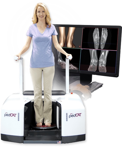
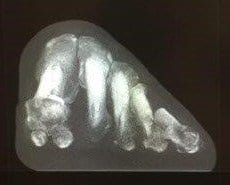
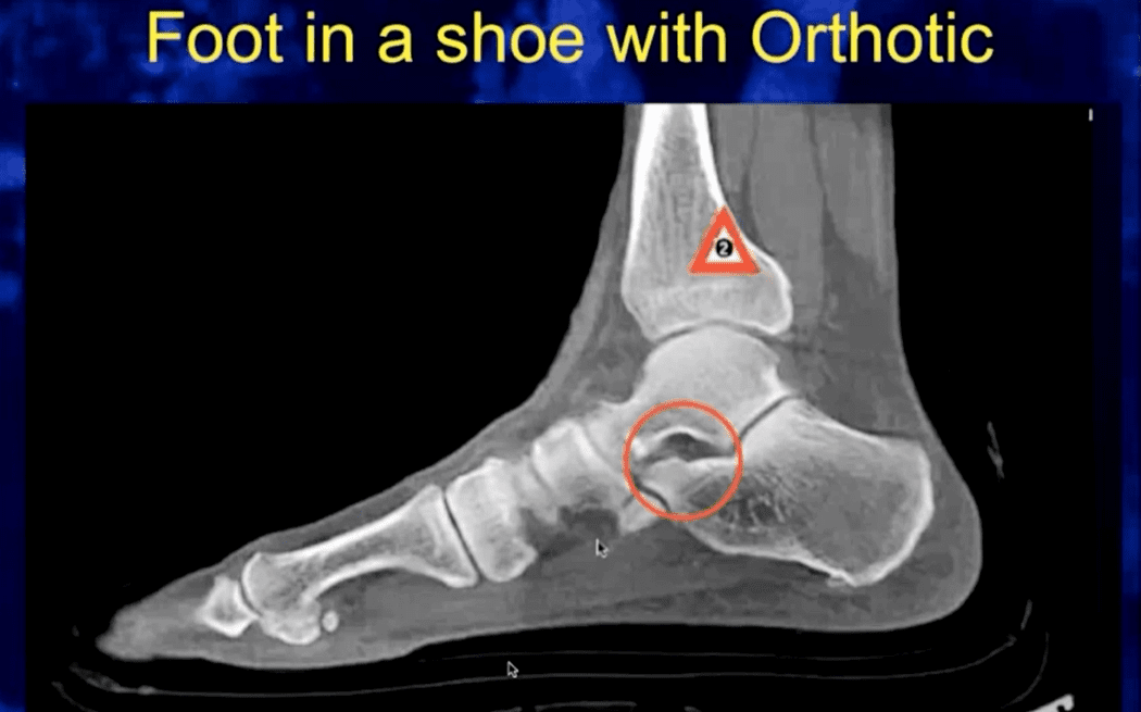
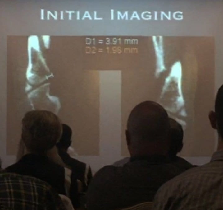
Dr. Michael Chin, DPM, presented how weight bearing CT imaging has changed how he evaluates cuboid subluxations at the American Academy of Podiatric Sports Medicine meeting held near the West Point Military Academy campus in early September, 2015. Dr. Chin began using the pedCAT in his office in February of this year. Not much research is out there on how to use plain radiographs to measure cuboid subluxations, Dr. Chin said in his lecture, titled, “Cuboid Syndrome…The Other Side of Heel Pain.” Dr. Chin has tested using a bilateral oblique projection to understand the cuboid/ metatarsal relationship, and has been able to observe a slide between the head of the fourth metatarsal and the head of the cuboid. An MRI could be ordered to see the condition of the peroneal tendon, but the study would be limited because the scan would not be weight bearing, he said.. A traditional CT scan would provide great visualization of the bone, but would provide no information on anatomic alignment. The pedCAT weight bearing CT imaging system is excellent for evaluating stress fractures, sesamoids, periosteal changes, or anything medullar, Dr. Chin said. Another benefit is he can measure the exact degree of subluxation between the cuboid and the fourth metatarsal head. Dr. Chin displayed pedCAT images depicting pre and post-reduction views of a cuboid subluxation. pedCAT scan of a pre-surgical patient with cuboid subluxation. Dr. Chin was able to reduce the subluxation to 2.18 mm. Dr. Chin practices at The Running Institute in Chicago.
Read more

AOFAS Annual Meeting – CurveBeam Symposium Recap

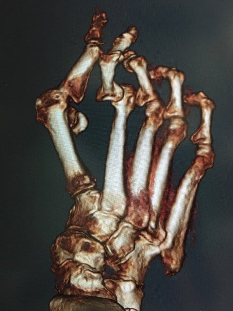
Weight bearing CT is a vital tool for determining the cause of inexplicable pain, and also for avoiding painful surgical complications. That was the takeaway message from a talk by Dr. Phinit Phisitkul, a clinical associate professor of orthopaedics at the University of Iowa. He shared some of his most interesting cases at a CurveBeam sponsored symposium held during the AOFAS Annual Meeting in Long Beach during the evening session. We’ve selected three of his cases to share on this blog: 18-year-old male with Noonan Syndrome & severe flat foot: The patient presented with an unusual amount of pain that was difficult to diagnose on plain X-Ray. A weight bearing CT scan revealed he had a severe deformity – a congenital vertical talus. He also had severe impingement. Vertical Talus - Weight Bearing CT Vertical Talus - Weight Bearing CT Impingement - weight bearing CT Impingement - weight bearing CT 58 year-old male with ankle arthritis: The patient presented with a lot of pain in the ankle joint. A weight bearing CT scan showed a subluxation of the ankle joint and dramatic impingement of the calcaneal fibula. Interestingly, the subtalar joint was in pristine condition. Dr. Phisitkul determined the patient was a good candidate for ankle replacement and hindfoot realignment, and that his subtalar joint could be spared. Calcaneal-fibular impingement and arthritis - Weight Bearing CT Calcaneal-fibular impingement and arthritis - Weight Bearing CT 41-year-old female with Hallux Valgus: A weight bearing CT scan revealed a bone spur on the patient’s first metatarsal head. If the doctor had done a normal release, the spur may have ended up pinching the sesamoid. Instead, he performed a lateral release and excised the bone spur.
Read more
Back To Top