Studies Point to Many Benefits of Weightbearing CT Scanning Technology
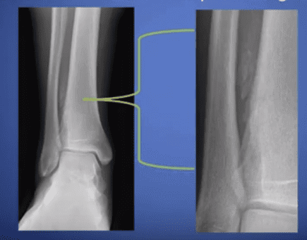
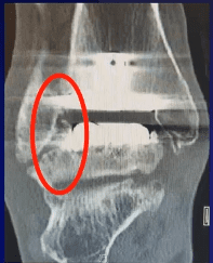
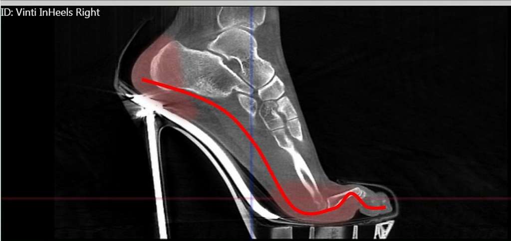
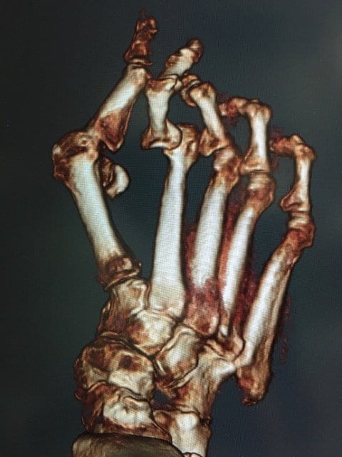
For patients whose lives are impacted by medical technology breakthroughs, there is a significant improvement in the quality of care their doctors are then able to provide. This is especially true when it comes to cone beam computed tomography (CT)…