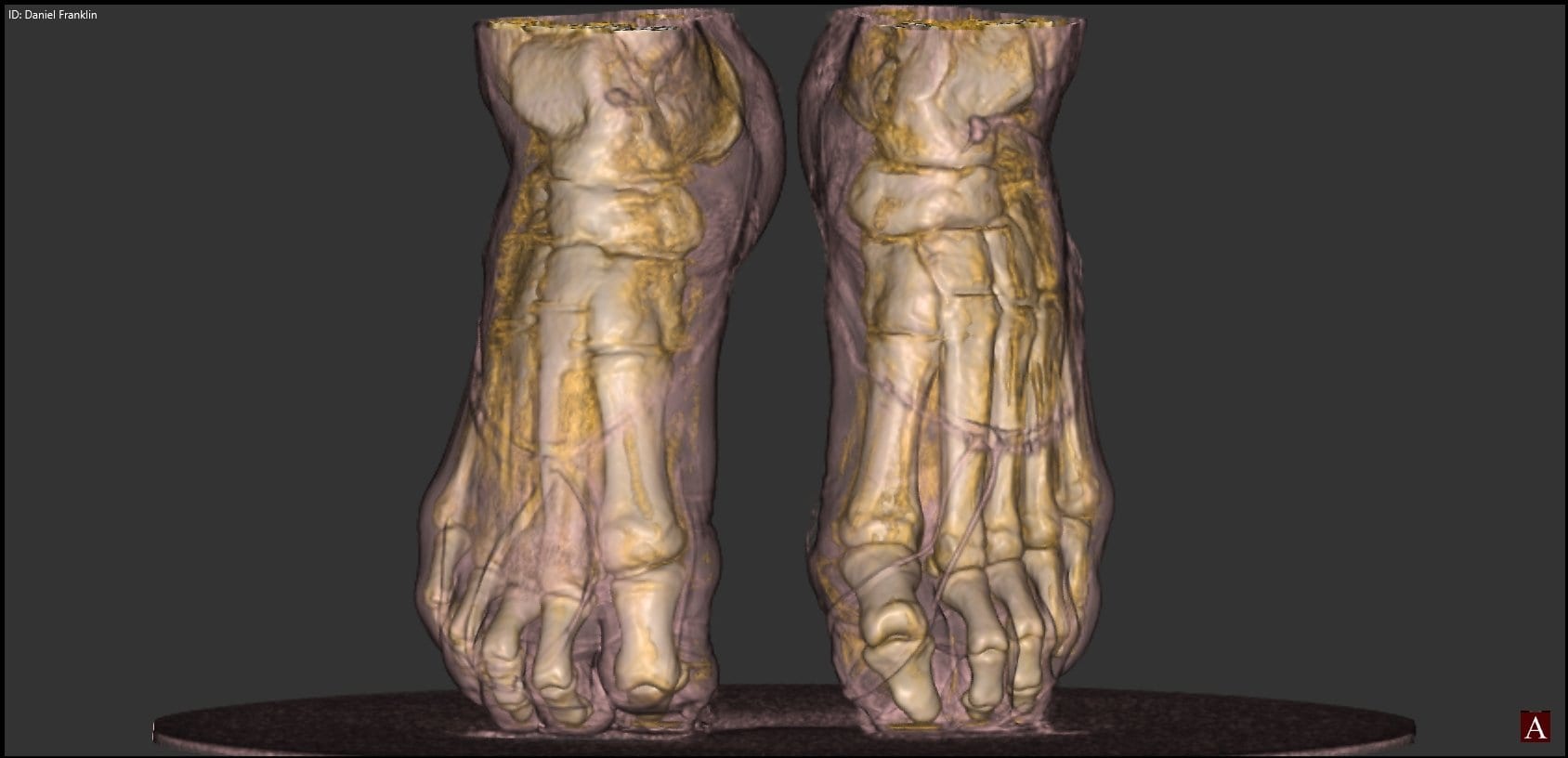
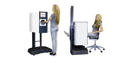
Chat with us, powered by LiveChat Skip to content

Case Study: pedCAT vs. Radiographs

A patient sought a second opinion for the cause of her medial foot and ankle pain. The treating physician used plain radiographs to diagnose her with posterior tibial tendon dysfunction/partial tear and degenerative joint disease of the 1st, 2nd, and 3rd tarsometatarsal joints. The treating physician also noted a “chip” of bone on the inside of the ankle. The treating physician planned on performing a flat foot reconstruction, a posterior tibial tendon repair, and a tarsometatarsal joint fusion. The physician performing the second opinion noted the patient’s discomfort over the medial ankle gutter was much more significant than over the posterior tibial tendon and the spring ligament. The patient had minimal discomfort through the tarsometatarsal joints. The physician performing the second opinion ordered a weight bearing pedCAT study to assess the midfoot DJD and to better evaluate the midtarsal joint and ankle joint. The pedCAT study clearly documented a degenerative process in the medial ankle gutter with a bony impingement. On secondary exam, the majority of the symptoms arose from the medial ankle gutter. If the flat foot reconstruction was performed as planned, the talus would have been dorsiflexed and the tibio-talar impingement would have been worsened. The pedCAT images helped prevent an un-necessary surgery, while directing the physician to the appropriate pathology.

Read more

pedCAT CubeVue: Automatic X-Ray Views

It takes less than a minute to scan a patient in the pedCAT, but that’s enough time for the pedCAT system to collect enough data to create a 3D reconstruction of the foot and ankle, as well as .3 mm slices in all three planes.

Read more

Distinguishing Categories of Medial Column Ligamentous Fracture

Weight bearing CT scans offer new, three-dimensional perspectives that reveal a better understanding of the biomechanics of flat foot. That was a finding on an academic poster on display at The American Orthotic and Prosthetic Association's National Assembly in Las Vegas (Sept. 4 - 9). "IRD, PTTD, Posterior Tib, Flat Foot, Pronation...these are all terms that various professions use to describe some sort of medial column ligamentous breakdown," according to the poster. "The purpose of this study is to better distinguish some types of pathomechanics of the foot. By categorizing and redefining them, it makes the description of pathology more useful for Orthotic Treatment. This was possible through the use of 3D rendering of the pedCAT machine and CurveBeam software." The study was conducted by Ian Engelman, M.S. CPO, and Harold Chamberlin, DPM. They looked at 3D renderings of the bony structures in flat feet, and came up with some new descriptions, as well as their possible ligamentous causes. The study concluded that while the exterior contours of the foot suggest certain bony pathologies, the 3D renderings provided by the pedCAT would allow for orthotists to better distinguish the pathomechanics of the foot and ankle. An unexpected result showed a moderately strong correlation (-0.68) between the vertical position of the navicular and medial displacement in a patient population with ranging degrees of medial column breakdown.

Read more

pedCAT: Weight Bearing CT Views of Lisfranc conditions

Lisfranc injuries can easily be missed on traditional radiographs. One research study tried to determine the level of accuracy when using plain radiographs to assess midfoot conditions. Their results confirmed a high incidence of missed diagnosis even by experienced observers.¹ “Radiographic evaluation is crucial in the diagnosis…Weight bearing views are more sensitive since tarso-metatarsal instability may be revealed…The threshold for cross sectional imaging such as multi-detector CT should be low,” according to the study. With the pedCAT, it is possible to get a weight bearing, three dimensional scan at the point of care. The tarso-metatarsal joint can be viewed without any superimposition from surrounding anatomy. Delaying a 3D scan can be detrimental to recovery. “The identification of Lisfranc joint injury can be difficult and is often not detected on initial presentation to the accident department. This is important because of the correlation between delay in treatment, particularly more than six months, and poor functional outcome,” the study reported. Click on the blog post title to see examples of weight bearing CT scans of Lisfranc injuries.

Read more

Webinar: Dr. Erik Nilssen – A New Paradigm in Foot & Ankle Diagnosis

“Patients come to my practice with an expectation of a higher level of care, that they’re getting the best. With the pedCAT, I am able to give them information immediately, and they leave feeling this was well worth the trip.” – Dr. Erik Nilssen Learn how the pedCAT has changed Dr. Nilssen’s clinical practice in a webinar hosted by Foot & Ankle International and Foot & Ankle Specialist. We hope you can join us! Click on the title of the blog post for the link to sign up.

Read more

pedCAT: Fracture & Fusion Assessments

X-rays of the foot and ankle may not always provide conclusive assessments for post-operative fusions such as of the tarso-metatarsal joint or hind foot joints. Similarly, the physician is often left guessing if a fracture has properly healed. “The pedCAT takes all the variability out, all the guesswork out of it,” Dr. Martin O’Malley, MD, an Associate Attending Orthopedic Surgeon at the Hospital for Special Surgery in New York, New York, said. Dr. O’Malley said pedCAT scans allow him to clearly determine if a fusion has healed more than 50 percent, and he decides when to ambulate his patient accordingly. “I let people walk on it earlier than before, and I keep them off longer than before,” Dr. O’Malley said. The pedCAT provides a three-dimensional view of fractures that changes the way O’Malley sees this common diagnosis. “These posterior pieces are often bigger than we thought were based on plain X-ray and they often travel all the way around the medial side as well, which we never thought they did,” Dr. O’Malley said. “You know, we thought it was an infrequent fracture, but now we see it routinely. Now most of my ankle fracture work, I’d say more than half the time, is through a posterior approach. For the first 15 to 18 years of my practice I would do medial/ lateral incisions. Now I’m going to the back of the ankle. And a lot of it is driven by the pedCAT.” pedCAT Stress Fracture Cross Section Click on the blog post title to see an example of a navicular stress fracture in a collegiate runner that had not healed at all after six weeks of casting. Were it not for the conclusive pedCAT scan, this patient would have been allowed to ambulate. Watch Dr. O’Malley talk more about the pedCAT here . To offer your patients state-of-the-art fracture & fusion assessment, consider a pedCAT for your practice.

Read more

pedCAT: Will it Fit?

The pedCAT has been deployed to a range of medical facilities – from large hospitals to single physician practices. Each site has its own unique requirements, and CurveBeam works with each customer to ensure the first weight bearing CT scanner dedicated to the foot & ankle meets all safety requirements. Because the pedCAT is an ultra low dose device, the shielding infrastructure required is similar to that of a plain X-Ray device. The pedCAT does not need to be in a lead lined room. Because of its compact size and relatively minimal required shielding, the pedCAT can easily fit into a practice with limited space. Click on the title of this post to see some examples.

Read more
Back To Top