AI-Driven Segmentation on Weight Bearing CT Introduction Accurate segmentation is critical for orthopedic workflows, including…
Case Study: WBCT for Visualizing the Cartilage and Menisci
Current Standard of Care
MRI is the current standard of care for non-invasive visualization of the cartilage and menisci. However, absence of weight bearing limits evaluation of these structures in a functional position.
Value Proposition CBCT Arthrogram
Weight Bearing CT arthrogram may be useful for assessment of menisci as well as tibiofemoral and patellofemoral cartilage in a functional stance.
With standing CT, specialists can obtain 3D imaging of the knees while they are under physiological load, similar to fixed-flexed or semi-flexed radiograph protocols.
Potential advantages over non-weight bearing MRI/MRA include:
• 3D measurements of the meniscal position and morphology while standing are more accurate.
• Visualizing pathology that is not detected in unloaded positions is now possible.
• Magnitude of muscle and external forces acting about the knee are better recreated during actual standing.
Positioning Advantages include:
• Ability to bear weight bilaterally in a functional position is important for accurate assessment of meniscal position, meniscal pathology under physiological loads, and assessment of cartilage thickness when loaded.
• Weight bearing CT arthrogram can be obtained in multiple knee flexion angles, while an MRI knee coil may permit imaging only with the knee in extension.
Financial Advantages include:
• A weight bearing CT arthrogram study is less expensive than MRI for patients and payers
• The 2022 Medicare National Payment amount for 73725 – MR Ang lwr ext w/ or w/o dye: $365.10
• The 2022 Medicare National Payment amount for 73701 – CT lower extremity w/ dye: $179.26

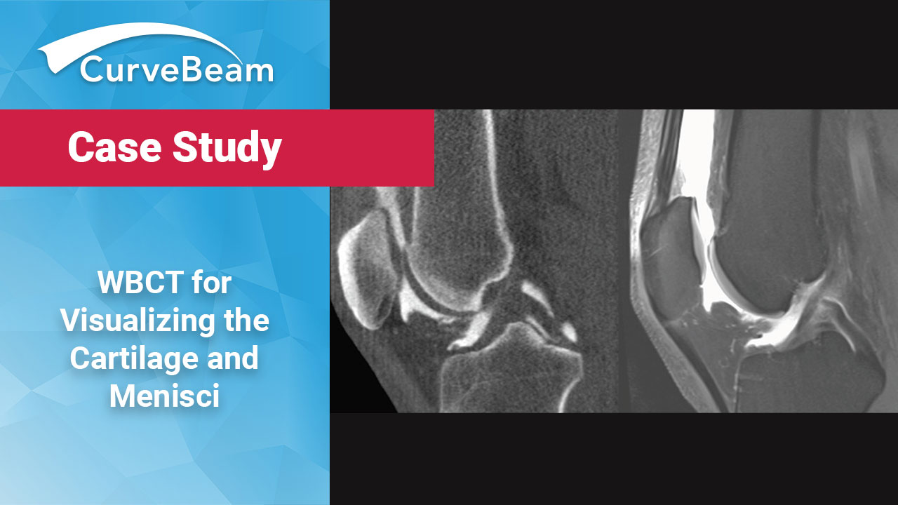
The sagittal reformatted weight bearing CT arthrogram (left) and MRI (right) both demonstrate outstanding delineation between cartilage and subchondral bone.
Technique for Weight Bearing CT Arthrogram
Inject a contrast mixture into the patient’s knee. Guide the patient to flex and extend the knee for 2-3 minutes while in a non-weight bearing position. Position the patient in a CurveBeam weight bearing CT scanner with the tips of the great toes, patellae, and the anterior superior iliac spines contacting the positioning frame that maintained these points coplanar to each other and the feet 10 degrees rotated. Acquire scans at 0.3mm voxel size. No recovery time is required after scanning.

Weight bearing CT arthrography (top) and corresponding MR arthrography (bottom) demonstrate outstanding delineation of the tibiofemoral and patellofemoral articular cartilage. Weight bearing CT shows better differentiation of cartilage and subchondral bone.

Weight bearing CT image (left) of a small tear in the tibial surface of the posterior horn of the medial meniscus and corresponding axial image (right) depicting location.
Segal NA. Advances in Visualization of Knee Cartilage with Standing Computed Tomography Arthrography. Poster presented at: Osteoarthritis Research Society International 2016 World Congress; March 31, 2016: Amsterdam, NL.
Related Posts


