AI-Driven Segmentation on Weight Bearing CT Introduction Accurate segmentation is critical for orthopedic workflows, including…
Utility of WBCT to Diagnose Syndesmotic Instability in Patients with Weber B Lateral Malleolar Fractures
Key Points
- WBCT is able to discern a stable from an unstable syndesmosis.
- WB X-Rays lack sensitivity to diagnose cases of subtle syndesmotic instability, especially in the presence of Weber B ankle fractures.
- WBCT syndesmosis volume measurements spanning from the tibial plafond to 5cm proximally had the largest area under the curve of 0.96 (sensitivity = 90%, specificity = 95%), which indicated an outstanding diagnostic test.
Historically, syndesmotic instability accompanied in patients with Weber B lateral malleolar ankle fractures is very difficult to diagnose. Most patients will undergo some form of intraoperative stress evaluation of the syndesmosis, though more information is generally needed on how to diagnose the fracture without medial clear space (MCS) distance on the initial radiographs.
A recent study by Dr. Rohan Bhimani et al. out of the Foot & Ankle Research and Innovation Laboratory (FARIL), Harvard Medical School, Massachusetts General Hospital in Boston, Massachusetts, found that utilizing both two-dimensional (2D) and three-dimensional (3D) Weight Bearing CT (WBCT) measurements, specifically syndesmotic joint volume measurements, were best suited to diagnose syndesmotic instability among patients with Weber B ankle fractures.
Materials and Methods
The study compared 23 patients treated for unilateral Weber B lateral malleolar ankle fractures with intraoperative syndesmotic instability requiring surgical fixation with a control group of 18 patients without intraoperative syndesmotic instability.
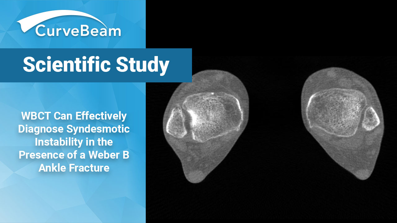
The following one-dimensional (1D) and 2D measurements were taken on both the injured and uninjured ankles: distal syndesmosis in the axial plane (including distance (1D) and area (2D) 1cm above the tibial plafond and included fibular rotation, tibiofibular distance measured at the anterior, middle, and posterior aspect of the distal tibiofibular articulation, and the syndesmotic area. Lateral malleolar fracture displacement was measured on the sagittal plane, and fibular shortening was measured on the coronal plane as represented by the distance between the fibular tip and the tibial plafond. MCS distance was measured on the mid-coronal CT slice for each patient as well.
For 3D measurements the following were recorded: distal tibiofibular articulation extending from the tibial plafond to 3cm and 5cm proximally. The MCS area (medial malleolus – talar dome), defined as the space between the medial malleolus and the talus delineated by two tangential lines adjoining the anterior margin and the posterior margin of the medial malleolus and the talus, and the lateral clear space (LCS) (lateral malleolus – talar dome) were measured in the axial plane slices (0.3mm standard thickness) starting from the distal most aspect of the medial tibiotalar joint to the superior extent of the talar dome. The LCS area, defined as the space between the lateral malleolus and the talus delineated by two tangential lines adjoining the anterior margin and the posterior margin of the lateral malleolus and the talus, was also measured in a similar fashion using the axial plane slices. The volume of the MCS and LCS was created and measured in the axial orientation using the following formula: Volume = [A1 x T] + [A2 x T] + [A3 x T]+. . .+[An x T], wherein A is the area in each slice and T is the slice thickness (in this study 0.3 mm).
Results
In patients with unilateral syndesmotic instability and Weber B ankle fractures, all WBCT measurements were significantly greater than the uninjured side, except MCS distance, syndesmotic area, and anterior and posterior tibiofibular distance. Additionally, syndesmosis volume spanning from the tibial plafond to 5cm proximally had the largest area under the curve of 0.96 (sensitivity = 90%, specificity = 95%), which indicated an outstanding diagnostic test, followed by syndesmosis volume up to 3cm proximally (area under the curve = 0.91; sensitivity = 90%; specificity – 90%). Apart from the MCS volume and distal fibular tip to tibial plafond distance, the control group showed no side-to-side difference in any parameter.
Conclusion
The study concluded that WBCT can effectively diagnose syndesmotic instability in the presence of a Weber B ankle fracture among patients with a symmetric MCS and who would otherwise not necessarily undergo surgical intervention. 3D volume measurements have a higher sensitivity than traditional distance measurements such as WB X-Ray, which lacks the sensitivity needed to diagnose subtle instability; or MRI, which readily identifies distal syndesmosis ligamentous injuries but not instability, and bony anatomy is often obscured in the acute trauma setting due to bony edema. Non-weight bearing conventional CT can be used to visualize syndesmotic alignment and bony detail but only does so unilaterally and without the patient under natural weight bearing circumstances. WBCT systems, such as CurveBeam’s HiRise, solves these issues by providing bilateral, high-resolution, 3D images which allow for an accurate diagnosis quickly and conveniently.
Researchers also noted that WBCT can be useful when the tibiotalar relationship appears to be intact but clinical concern for syndesmotic instability exists, which may necessitate surgical stabilization. Additional studies are necessary to evaluate the degree of syndesmotic asymmetry that clinically justifies repair and the effect of associated fractures of the posterior malleolus on such measurements.
To read the full study click here.
Click here to watch a webinar about a study out of Denmark that showed WBCT of patients with potentially unstable Weber B fractures found additional clinical findings that the initial X-Ray exams missed.
Related Posts


