
AI-Driven Segmentation on Weight Bearing CT Introduction Accurate segmentation is critical for orthopedic workflows, including…
WBCT + Coverage Mapping Finds Significant Subluxation of the TTJ in PCFD Patients
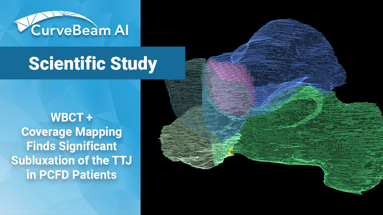
Key Points
- Weight bearing CT (WBCT) combined with coverage mapping (CM) and 3D distance mapping (DM) techniques may help improve understanding of bone positioning and interactions through the transverse tarsal joint (TTJ) in progressive collapsing foot deformity (PCFD).
- Patients with PCFD presented with significant coverage joint changes in the TTJ as compared to controls.
- 3D mapping, specifically related to plantarflexion and subluxation may be of particular value in diagnosing, staging, and estimating treatment impacts in PCFD.
Researchers at the University of Iowa applied joint coverage & distance mapping technology to weight bearing CT scans in an attempt to understand what happens to the transverse tarsal joint (TTJ) in patients who have progressive collapsing foot disorder (PCFD). Their results may provide much needed data to guide disease management and could lead to earlier diagnosis and better treatment.
Most of the pathology associated with PCFD is found in the peritalar subluxation of the TTJ. Little is known about the relationship of these joints, both normal and pathological. Little is also known about whether abduction of the midfoot, medial arch collapse, and forefoot varus have a substantial or minor contribution from the talonavicular and the calcaneocuboid joint.
Previous WBCT studies found that the amount of subluxation at the posterior and middle facets is correlated with PCFD diagnosis and severity.
University of Iowa researchers analyzed the subtalar joint in 20 stage I flexible PCFD patients and 20 controls using 3D distance maps (DMs) and coverage maps (CM) on WBCT images.
No significant differences were found in the DM measurements between control and PCFD patients. However, when it came to coverage mapping, a significant difference was observed. In PCFD patients:
- There was a significant decrease in coverage on medial talar head and plantarmedial regions of the calcanelcuboid interface.
- Subluxation occurred on both the talar head and calcaneocuboid facet. Significant subluxation was noted on the medial side of the talar head in patients with PCFD, especially on its plantar aspects. Parrellely, the lateral regions on the talar head had increased coverage when compared to controls.
- These results eliminated pure plantarflexion as the cause of subluxation and instead pointed to medial abduction and external rotation of the navicular being the root cause.
The information provided by the 3D mapping, specifically related to plantarflexion and subluxation may be of particular value when diagnosing, staging, and estimating treatment impacts in PCFD.
It was concluded that further research is needed to increase reliability through automation and to continue the search for more complete understanding of physiological bone congruence and changes associated with collapse in order to halt articular degeneration in PCFD.
To read the full study please click here.
Related Posts


