Orthopedic decision-making depends on accurate representation of anatomy—particularly when joint alignment and bone relationships change…
Cone Beam CT Helps Visualize Loose Bodies
A loose body is a bone or cartilage fragment that has chipped off inside a joint. If left in place, a loose body can damage a joint surface, cause pain, and restrict movement.
A 2D radiograph is typically the first test performed when looking for a loose body, but overlapping bone may obscure loose bodies. Cone Beam CT imaging provides a 3D image of the foot & ankle at a dose comparable to 2D imaging.
Dr. Albert Armstrong, DPM, MS, BSRS, Professor of Radiology and Medical Director of Advanced Imaging at the Barry University School of Podiatric Medicine, shared such a case in a recent FOOTInnovate lecture.
A 38-year-old male patient presented with pain in his left foot, mainly in the great toe. He said his toe felt stiff and would often “lock up.”
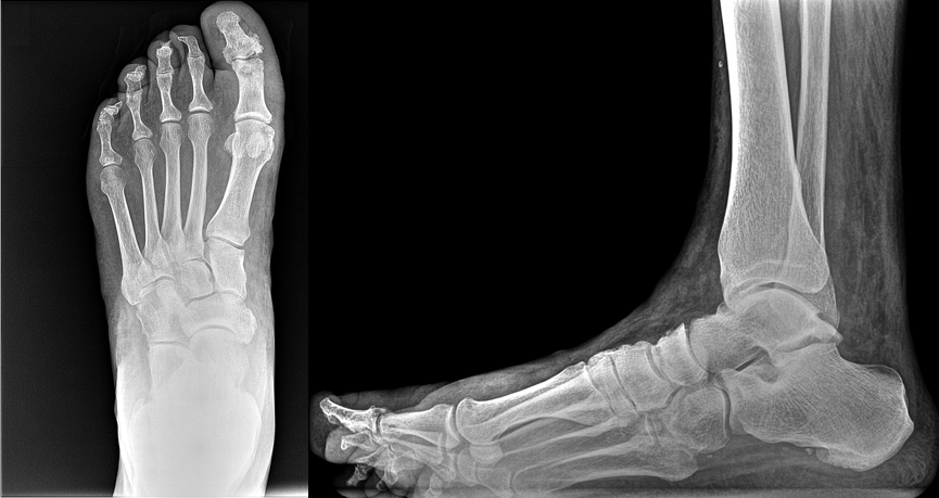
A cone beam CT scan was ordered as a follow-up exam. Because the podiatric clinic at Barry University has a CurveBeam pedCAT on site, the patient was able to get the scan immediately.
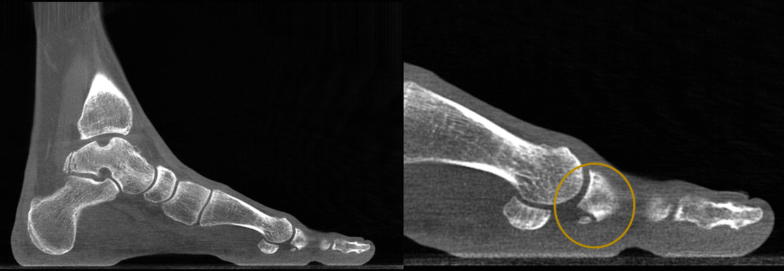
The cone beam CT scan revealed osteophyte formations on the 1st metatarsophalangeal joint, and a resulting loose body.
According to a study published in the European Journal of Radiology in 2017, that because of its low radiation dose, cone beam CT “can be used in current practice as a replacement or supplement to radiographs.”
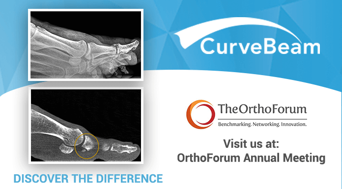
Are you attending the OrthoForum Annual Meeting in Phoenix? Be sure to visit the CurveBeam exhibit to learn more about our imaging solutions.

