
Cone Beam CT Helps Visualize Degenerating TMT Joints
Midfoot arthritis is a challenging problem that causes foot pain and can impede daily activity. Surgery, specifically midfoot arthrodesis, is considered when initial conservative management fails. Arthrodesis should be limited to the symptomatic joints, but it is often difficult to determine which joints are causing the symptoms. Precise anatomic preoperative diagnosis is essential (1).
Cone Beam CT imaging can assist surgeons in understanding complex forefoot deformities and devising the appropriate surgical plan.
For example, in the case below, a patient presented with forefoot pain and was a candidate for surgical revision after X-Ray exams revealed a forefoot deformity.

Based off of the X-Ray images alone, the treating doctor would have performed a scarf osteotomy and Weil procedures on the 2nd and 3rd metatarsals.
A follow-up weight bearing cone beam CT scan via a CurveBeam pedCAT was ordered.
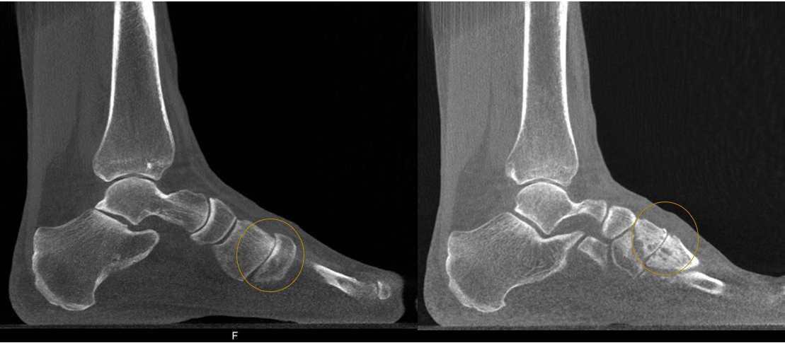
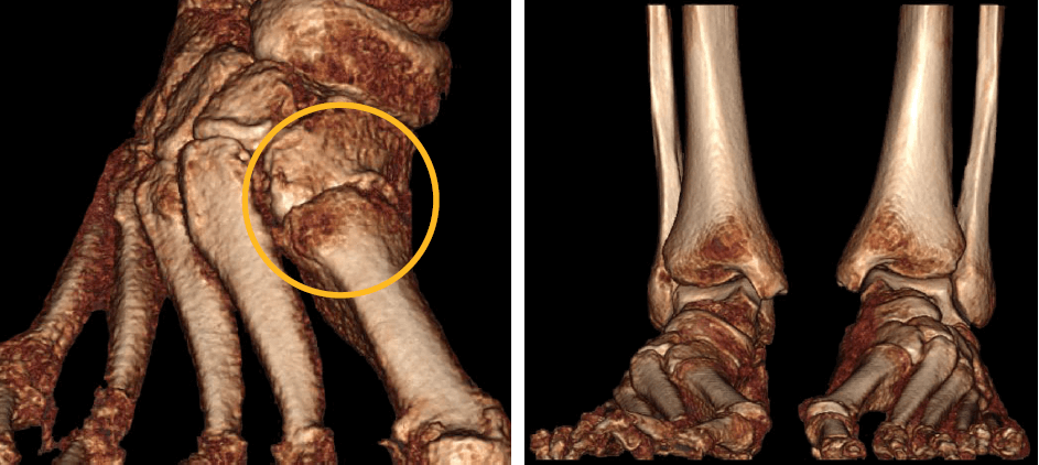
Based on the weight bearing CT scan, the surgical plan was revised to a Lapidus bunionectomy and a 2nd and 3rd tarsometarsal joint arthrodesis.
Will you be attending the AAOS Annual Meeting in Orlando? Visit CurveBeam at Booth #2909 to learn more about weight bearing cone beam CT imaging.

(1) Verhoeven N, Vandeputte G. Midfoot arthritis: diagnosis and treatment. Foot Ankle Surg. 2012;18(4):255–262. doi:10.1016/j.fas.2012.04.004
Related Posts