SSR – Cone Beam CT Helps Visualize Fusion Healing
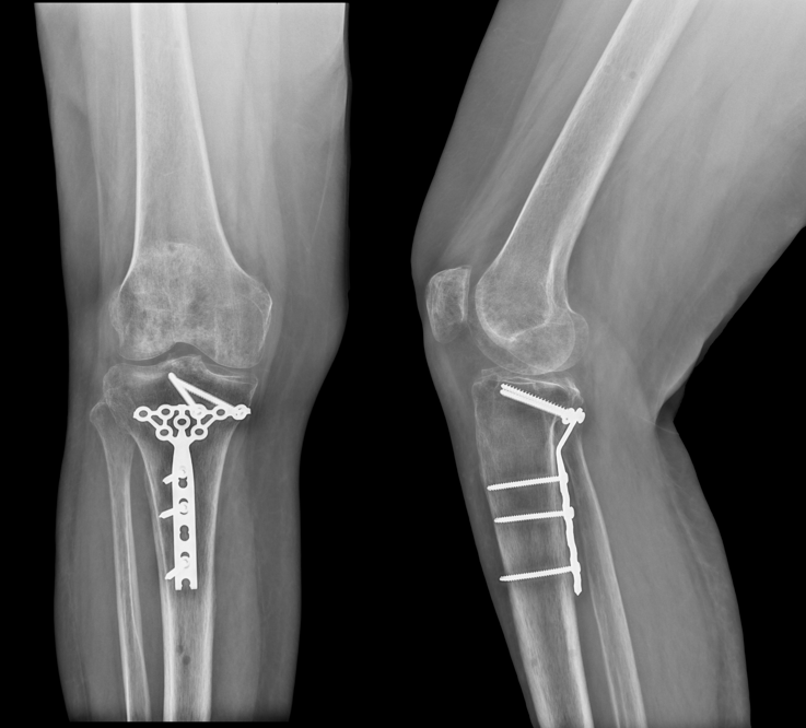
X-rays of knee may not always provide accurate post-operative assessments. For example, in the case below, post-operative X-Rays suggested a posterior shear tibial plateau fracture had been sufficiently healed.
However, a CurveBeam LineUP weight bearing CT exam of the same patient showed a portion of the fracture had not fused as desired.
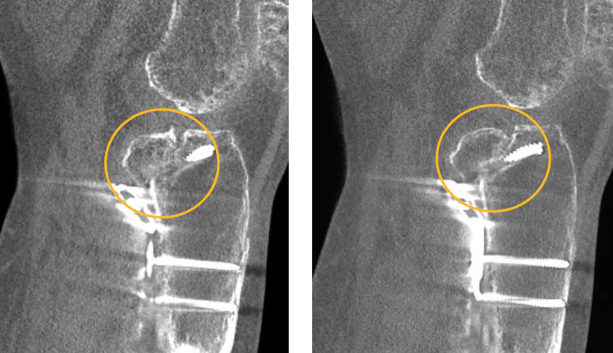
The LineUP provides three-dimensional views of fractures. The treating physician for this case, Dr. Blake Moore, said weight bearing CT has been the most critical addition to his practice since he began it, adding that it “greatly enhances the level of care and sophistication of preoperative planning.”
Are you attending the SSR Annual Meeting? Be sure to visit CurveBeam’s exhibit to learn more about weight bearing CT imaging and how it is revolutionizing orthopedic medicine.

Related Posts