Syndesmosis A syndesmotic ankle sprain, also known as high ankle sprain, is an injury to…

Mayo Clinic Case Report: WBCT Reveals Tibial Plafond Stress Fracture in Competitive Athlete
Key Points:
- Stress fractures should always be considered in athletes with ankle pain.
- This is the first published case in which weight bearing CT (WBCT) was used in diagnosis and surgical planning of a stress fracture.
In the sport of track and field, jumping events have proven to be some of the most injury prone. Pole vaulting is particularly susceptible to foot and ankle injuries. Stress fractures are relatively common in athletes; however, fractures of the anterior tibial plafond and medial malleolus are rare and even more so in jumping-based sport athletes. Only one case has been reported since 1990 in a professional basketball player, to the best of researcher’s knowledge. This case, published by radiologists and orthopedic surgeons at Mayo Clinic Arizona in the Journal of Bone and Joint Surgery is important for healthcare providers to understand the factors contributing to this pathology and keep the diagnosis in mind when evaluating an athlete presenting with ankle or foot pain, as delayed treatment increases the risk of future complications.
Case Report
A 25-year-old male competitive pole vaulter sought an evaluation after 5 months of right ankle pain that had been limiting his training. He said the pain worsened with launching and landing motions and improved with rest. Physical examination revealed no significant limitations to passive range of motion and found no obvious deformities, swelling or ecchymoses. Their right ankle was neurovascularly intact with good strength, and they demonstrated a normal gait pattern.
Initial X-Rays showed small osteophytes at the medial tibiotalar articulation with a small intra-articular body raising the possibility of anterior impingement.
Conventional medical CT (MDCT) imaging can detect cortical fracture lines but is not considered an appropriate initial test by the American College of Radiology Appropriateness Criteria. WBCT imparts less radiation dose than MDCT and “in instances where physical examinations and primary imaging tests are unrevealing, WBCT may be helpful in better visualizing bony abnormalities and defining anatomy for preoperative planning,” according to the case report authors.
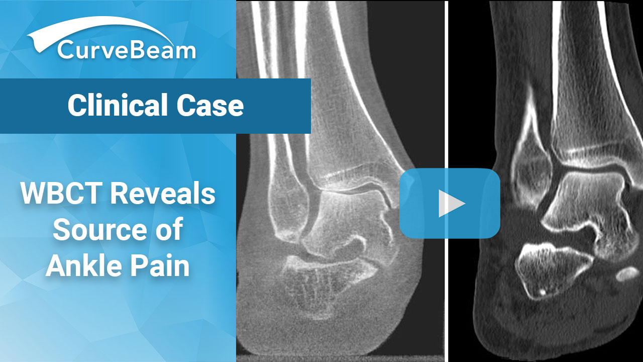
WBCT imaging of the ankle with maximal dorsiflexion was ordered, and scans showed severe narrowing with focal bone-on-bone apposition along the anteromedial aspect of the right tibiotalar joint. A nondisplaced stress fracture was also identified at the anteromedial tibial plafond at the level of the osteophytes.
“The ability to obtain CT images of the ankle dorsiflexed during weight bearing was important for confirming the diagnosis of an ankle osseous impingement with a tibial stress fracture and helped guide surgical management in our patient,” the authors wrote.
Magnetic resonance imaging (MRI) without contrast confirmed the incomplete fracture of the anterior distal tibia with extension into the articular surface and surrounding bone marrow edema.
The patient underwent a successful right ankle arthroscopy with anteromedial ankle osteophyte resection and loose body removal. Postoperative X-Rays showed successful resection of anterior tibial and talar osteophytes with no further imaging evidence of impingement.
To read the full case study click here.


