It is feasible for a single-practitioner podiatry practice to add weight bearing CT (WBCT) imaging and realize economical…
WBCT Indication Series: Charcot Arthropathy
Charcot Arthropathy
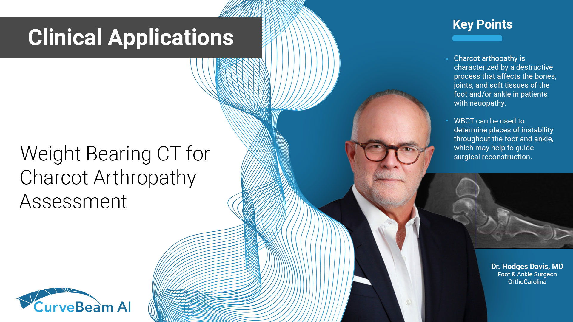
Charcot arthropathy is characterized by a destructive process that affects the bones, joints, and soft tissues of the foot and/or ankle in patients with neuropathy.
A weight bearing CT scan can:
- Assist in early detection and aid in an informed plan for early intervention, reducing complication risks1.
- Be used to monitor progression of deformity and help determine appropriate stage in which to surgically intervene, with the goal of preventing ulceration and infection.
- Provide insight into sites of instability.
- Determine areas of bony prominence that may lead to ulceration.
A WBCT scan provide valuable information regarding sites of Charcot arthropathy. Additionally, in the early stages of Charcot (Eichenholtz stages 0 and 1), a WBCT scan at regular intervals may be used to track progression and instability of the disease process.

X-Ray

Cone Beam CT
")
WBCT can be used to determine places of instability throughout the foot and ankle, which may help to guide surgical reconstruction. Additionally, WBCT scans can provide information about the size and quality of remaining bone stock in the foot. WBCT scans also can identify sites of bony prominence to plan exostectomies and prevent ulceration.
WBCT can improve surgical indications by better evaluating the severity of midfoot collapse, which has been shown to be associated with ulceration2.
Postoperative Assessment
For postoperative assessment of operative treatment of Charcot arthropathy, WBCT can:
- Accurately assess bony bridging across fusion sites3.
- Provide a three-dimensional analysis of deformity correction.
- Help to understand additional sites of collapse or instability.
Progression of Deformity
75 yo female with Charcot deformity that developed over the past one year. No reported neuropathy prior to diagnosis. Already on insulin with high A1c. History of dorsal foot ulcers. Presented with unstable varus ankle deformity.

Non-WB radiographs could not determine instability or severity of midfoot collapse; surgical intervention postponed.

One year later, deformity had significantly progressed, but the remaining bone stock was unclear from plain radiographs.

A preoperative WBCT scan showed the positioning and remaining bone of the tibia, calcaneus, & talus. Based on the WBCT scan, the decision to perform a tibiocalcaneal fusion with a hindfoot nail and frame was made.
Postoperative Assessment
65 yo male with a history of left midfoot ulcers. The patient was diagnosed with Charcot arthropathy.
WB radiographs of the left foot demonstrated Charcot arthropathy. However, evaluation of the midfoot, talonavicular, and subtalar joint was limited on these two-dimensional images.


Due to uncertainty regarding instability and position of the talus, a WBCT scan was obtained. The WBCT demonstrated lateral subluxation of the calcaneus under the talus. It also demonstrated bone loss and the position of the talonavicular joint. The WBCT suggested that the tibiotalar joint was salvageable.
The WBCT scan allowed preoperative surgical planning for deformity correction. The surgeon elected to perform a triple arthrodesis using a

midfoot nail to stabilize the medial column and talonavicular joint, a beam across the subalar joint, and a beam from the calcaneus into the midfoot.

Dr. Hodges Davis, MD
Dr. Hodges Davis, an OrthoCarolina Foot and Ankle Surgeon, has over 37 years of medical experience. His clinical interests include ankle arthritis, neuropathic disease, forefoot reconstruction, and deformity.
(1) Wukich DK, Sung W, Wipf SA, Armstrong DG. The consequences of complacency: managing the effects of unrecognized Charcot feet. Diabet Med. 2011 Feb;28(2):195-8. doi: 10.1111/j.1464-5491.2010.03141.x. PMID: 21219429.
(2) Radiographic Analysis of Diabetic Midfoot Charcot Neuroarthropathy With and Without Midfoot Ulceration. Dane K. Wukich, MD1,2, Katherine M. Raspovic, DPM3, Kimberlee B. Hobizal, DPM2, and Bedda Rosario, PhD4. Foot and Ankle International. 2014. November 35(11): 1108-1115.
(3) Armstrong, A. V. (2016, October). Current Concepts With Weightbearing CT. Hmpgloballearningnetwork.com. Retrieved March 30, 2023, from https://www.hmpgloballearningnetwork.com/site/podiatry/current-concepts-weightbearing-ct
Related Posts