It is feasible for a single-practitioner podiatry practice to add weight bearing CT (WBCT) imaging and realize economical…

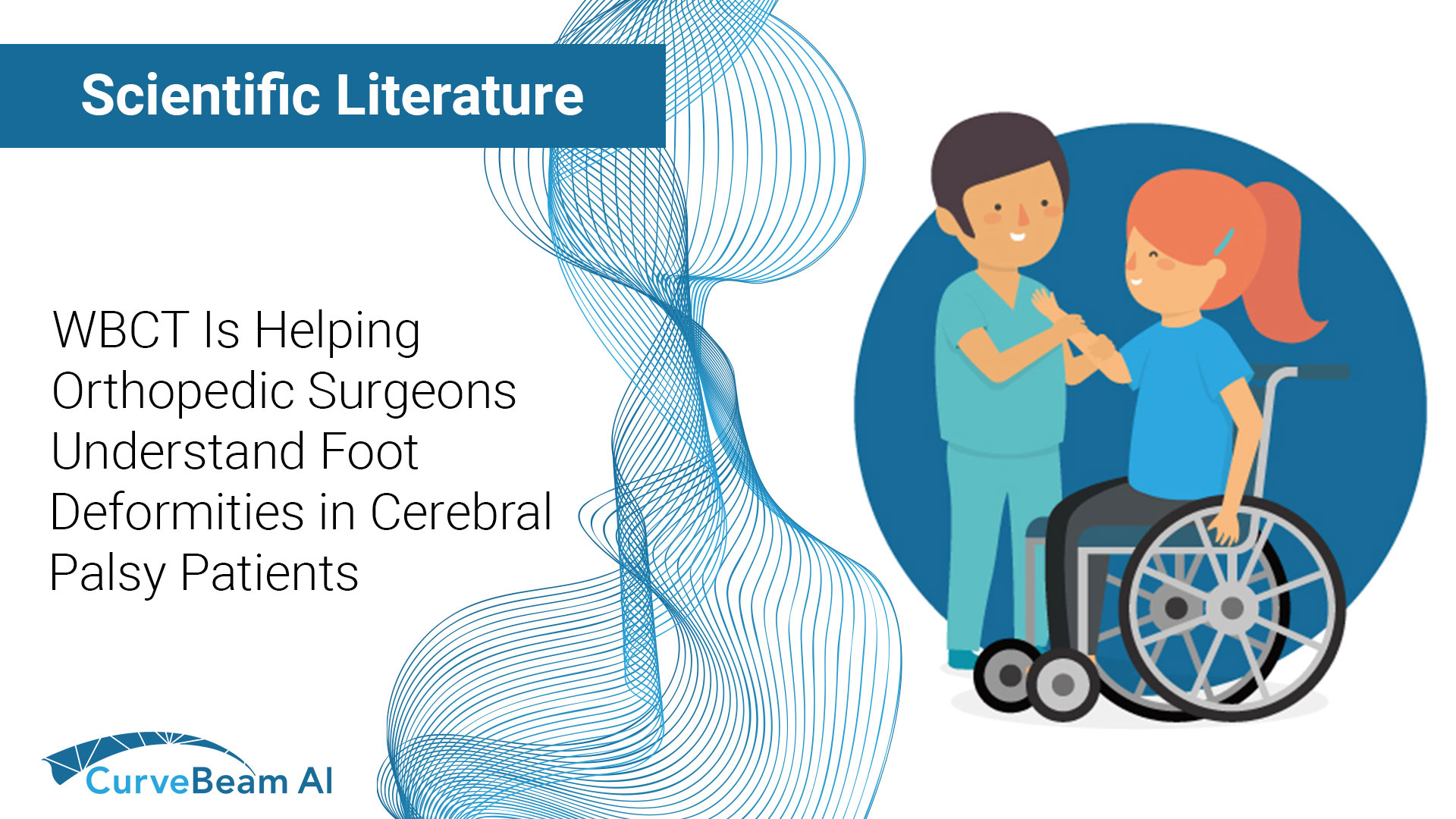
WBCT is Helping Orthopedic Surgeons Understand Foot Deformities in Cerebral Palsy Patients
Weight bearing CT scans are helping us understand pediatric foot deformities better than ever before.
Did you know 90% of children with cerebral palsy develop a foot deformity? Relative position and orientation of the talus, calcaneus, and navicular bones are key in diagnosis and treatment of these deformities, but X-Rays and 3D gait analysis are limited, especially to assess talar position.
Researchers in the Netherlands have found weight bearing CT could provide clinically valuable pre- and post-operative information for these patients. They used 3D bone analysis software to compare 14 cerebral palsy patient feet to 20 controls. Compared to controls, the cerebral palsy patients had much more variability. For the young patients in this cohort, calcaneus and navicular position in relation to the talus tended to be abnormal.
Researchers said 3D visualization could help classify and grade the severity of these foot deformities and serve as a visual guide for pre-operative planning. Cutting guides could also be designed using this data.
Weight bearing CT is helping orthopedic surgeons treat their most precious patients.
Will you be at IPOS in Orlando this December? Visit CurveBeam AI to learn how weight bearing CT might be the right solution for your practice.
Access the full study here: https://pubmed.ncbi.nlm.nih.gov/36585514/


