It is feasible for a single-practitioner podiatry practice to add weight bearing CT (WBCT) imaging and realize economical…
WBCT Indications Series: PCFD
Progressive Collapsing Foot Deformity
Progressive Collapsing Foot Deformity (PCFD), previously known as adult-acquired flatfoot deformity, is a multifactorial disorder characterized by concurrent multiplanar bony deformities. These deformities include, but are not limited to, hindfoot valgus, flattening of the medial longitudinal arch, peritalar subluxation, and midfoot abduction.
A weight bearing CT scan can:
- Provide an assessment of important anatomical markers of pronounced hindfoot deformity and peritalar subluxation (PTS), difficult to visualize on conventional two-dimensional radiographs1.
- Allow for accurate evaluation of subtalar joint subluxation as well as sinus tarsi and subfibular impingement2.
- Allow optimized and reliable evaluation of three-dimensional bone alignment under physiological standing loading3.
- Assist in planning operative reconstruction by determining the need for osteotomies and fusions as well as better estimating the amount of surgical correction necessary to properly realign the foot4.
Diagnosis
Because PCFD is a complex and dynamic three-dimensional deformity, WBCT offers several unique advantages including an improved spatial resolution. The multiplanar three-dimensional evaluation minimizes rotational and positional bias and bone overlap1.
In addition, WBCT better quantifies the structural deformity of PCFD compared to conventional radiography and non-weight bearing CT images1.
Treatment Planning
The high reliability and reproducibility of WBCT images in the three-dimensional evaluation of the PCFD can help the orthopedic surgeon to determine the amount of correction needed to achieve the best realignment of the patient’s foot1.
It can also be used to effectively assess joint morphology5, demonstrating a standard baseline threshold of normal anatomy, with implications for surgical planning in hindfoot reconstructive surgery.
Postoperative Assessment
For postoperative evaluation of operative treatment of PCFD, a WBCT can:
- Accurately assess the adequacy of correction4.
- Evaluate and follow postoperative deformity correction over time.
- Accurately assess healing of hindfoot and midfoot joint fusions6.
- Accurately assess subtalar and subfibular impingement2.
Evaluation of the Progressive Collapsing Foot Deformity
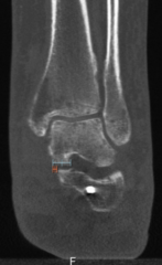
33 yo female who sprained her left ankle while dancing at a party and felt a pop on the medial side of her ankle. Although she has had flat feet since childhood, she has noted the progressive collapse of her medial arch in comparison with the contralateral side.
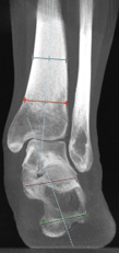
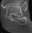
The WBCT scan was able to demonstrate subluxation at the medial facet of the subtalar joint and hindfoot valgus.
Post-Op Assessment
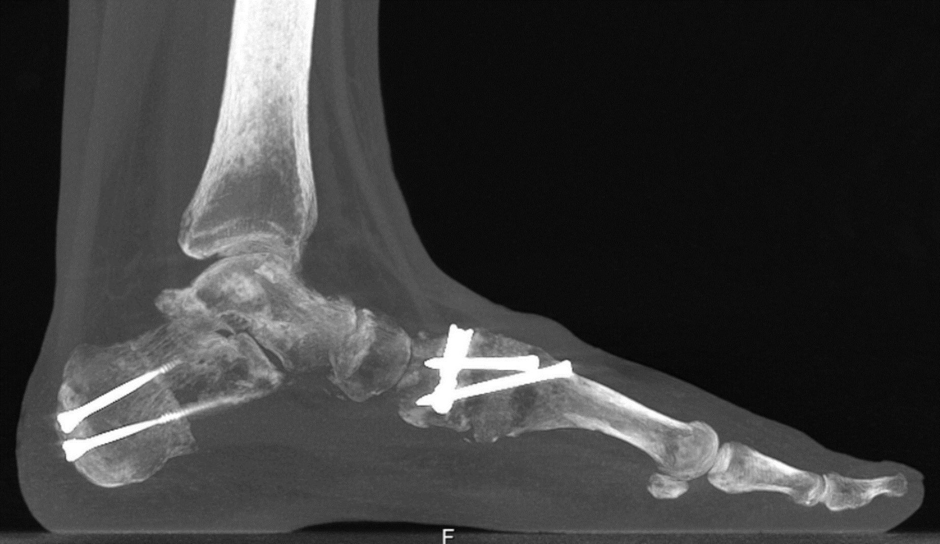
45 yo female was completing a kickboxing drill and felt sudden pain in her left foot while kicking an object. She has had flat feet since childhood. She underwent fusion of the first tarsometatarsal (TMT) joint with a Cotton osteotomy and calcaneus osteotomy to address her instability. Postoperative WBCT assisted in the
visualization of adequate reduction of the peritalar subluxation and allowed assessment of healing of the osteotomies and fusions.
Click Here to get direct links to the latest studies using WBCT scans.

Dr. Cesar de Cesar Netto, MD
Dr. Cesar de Cesar Netto, an University of Duke Health Foot and Ankle Surgeon, has studied multiple pathologies of the foot and ankle, with focus on flatfoot deformity, Achilles tendinopathy and advanced imaging of the foot and ankle.
(1) de Cesar Netto C, Myerson MS, Day J, Ellis SJ, Hintermann B, Johnson JE, Sangeorzan BJ, Schon LC, Thordarson DB, Deland JT. Consensus for the Use of Weightbearing CT in the Assessment of Progressive Collapsing Foot Deformity. Foot Ankle Int. 2020 Oct;41(10):1277-1282. doi: 10.1177/1071100720950734. Epub 2020 Aug 27. PMID: 32851880.
(2) Jeng CL, Rutherford T, Hull MG, Cerrato RA, Campbell JT. Assessment of Bony Subfibular Impingement in Flatfoot Patients Using Weight-Bearing CT Scans. Foot Ankle Int. 2019 Feb;40(2):152-158. doi: 10.1177/1071100718804510. Epub 2018 Oct 8. PMID: 30293451.
(3) Dibbern KN, Li S, Vivtcharenko V, Auch E, Lintz F, Ellis SJ, Femino JE, de Cesar Netto C. Three-Dimensional Distance and Coverage Maps in the Assessment of Peritalar Subluxation in Progressive Collapsing Foot Deformity. Foot Ankle Int. 2021 Jun;42(6):757-767. doi: 10.1177/1071100720983227. Epub 2021 Jan 27. PMID: 33504217.
(4) Rojas EO, Barbachan Mansur NS, Dibbern K, Lalevee M, Auch E, Schmidt E, Vivtcharenko V, Li S, Phisitkul P, Femino J, de Cesar Netto C. Weightbearing Computed Tomography for Assessment of Foot and Ankle Deformities: The Iowa Experience. Iowa Orthop J. 2021;41(1):111-119. PMID: 34552412; PMCID: PMC8259196.
(5) Ortolani, M., Leardini, A., Pavani, C. et al. Angular and linear measurements of adult flexible flatfoot via weight-bearing CT scans and 3D bone reconstruction tools. Sci Rep 11, 16139 (2021). https://doi.org/10.1038/s41598-021-95708-x
(6) Steadman J, Sripanich Y, Rungprai C, Mills MK, Saltzman CL, Barg A. Comparative assessment of midfoot osteoarthritis diagnostic sensitivity using weightbearing computed tomography vs weightbearing plain radiography. Eur J Radiol. 2021 Jan;134:109419. doi: 10.1016/j.ejrad.2020.109419. Epub 2020 Nov 21. PMID: 33259992.

