At CurveBeam AI, we often talk about the clinical and workflow advantages of weight-bearing CT,…

Charcot Foot: Dx w/ WBCT Imaging

 By Dr. David J. Soomekh, DPM, FACFAS
Guest Contributor
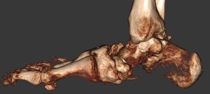
Charcot foot is a complex foot deformity involving subluxations and dislocations and fractures of the foot. It is most commonly seen in patients with uncontrolled Diabetes or other neurological deficiencies. When the nerves to the foot and ankle are not working properly, the blood vessels may increase blood flow to the bones, causing the bones and joints to get weaker, leading to collapse of the joints and bones. The foot may become flat and deformed and develop bone prominences that can become painful and cause open wounds.
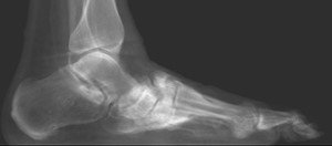
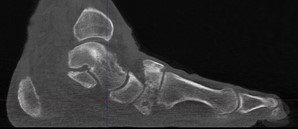
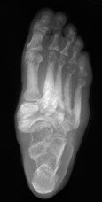
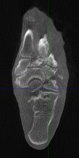
Traditional X-Ray can not give the physician the amount of information needed to access the complex midfoot and rearfoot joints in early stages of Charcot. MRI images will show inflammation in the bones, yet make it more difficult to address the spatial orientation of the bones and joints as compared to the weight bearing CT scan.
It is critical to diagnose Charcot deformity in its early stages in order to reduce the chance of significant collapse or fractures. Early diagnosis is best done with CT imaging. The pedCAT weight bearing in office CT system gives the treating physician a 3-dimensional view of the orientation of the joints. It will also show the difference in the stages of Charcot foot. This is important as each stage is treated differently.
Related Posts