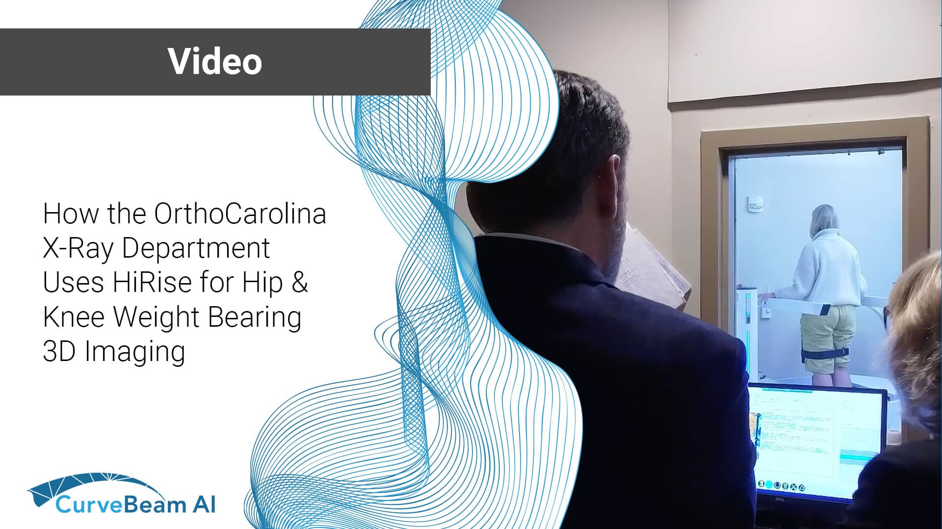
At CurveBeam AI, we often talk about the clinical and workflow advantages of weight-bearing CT,…
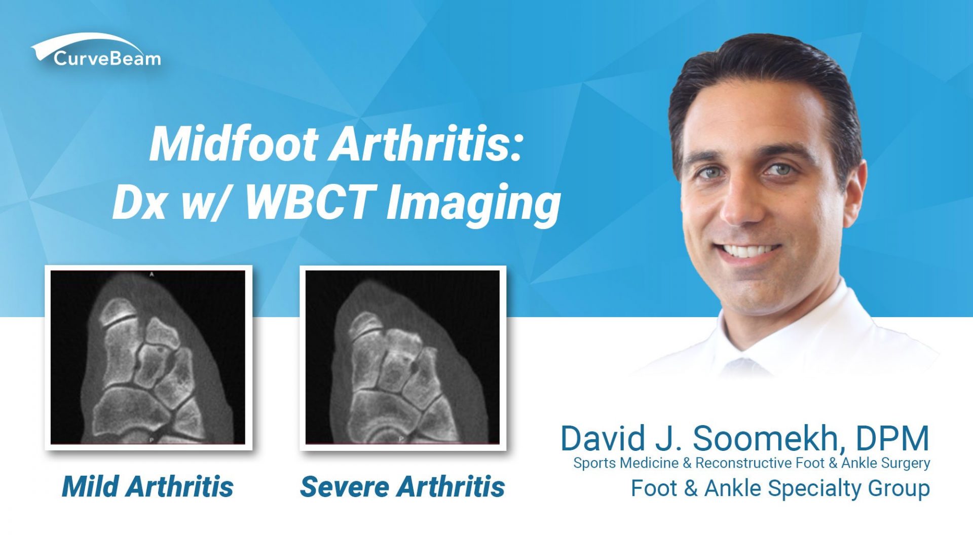
Midfoot Arthritis: Dx w/ WBCT Imaging

 By Dr. David J. Soomekh, DPM, FACFAS
Guest Contributor
Osteoarthritis is the loss of cartilage between 2 bones. This kind of arthritis can occur in any joint of the body. The foot and ankle are uniquely susceptible due to the significant stresses that can occur in the foot and ankle joints.
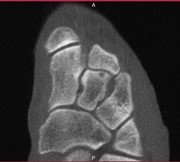
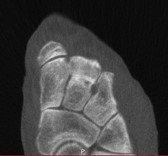
Behind the 1st, 2nd and 3rd metatarsals (the long bones of the foot) are the cuneiforms and the joints between them. These joints can become damaged over time from a micro or as a result of significant trauma like a fracture through the joint. Once the damage begins, the cartilage between the bones will progressively wear down until there is no barrier between the bone surfaces. This will cause pain and difficulty walking.
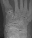
Treatment of osteoarthritis of the midfoot in the early stages is critical to avoiding chronic pain or surgery. Early diagnosis is the key. Traditional X-Ray radiographs of the midfoot are limited in their ability to show the early stages of the arthritis. The orientation of the bones and joints are such that these specific joints can be obscured on the film and not well visualized. MRI imaging of the foot can be more valuable than x-rays, yet does not offer the ability to visualize the relationship of the bones while the foot is in weight bearing.
Using in-office weight-bearing CT to evaluate arthritis gives the doctor significantly more information. This allows the surgeon to evaluate the position of the bones with weight and if there is any contribution to the degeneration of the joints more anatomically. With the ability to see the degree of damage to the joint and the amount of bone spur formation in three dimensions, conservative or surgical planning can be more precise. This allows the surgeon to better prepare the patient for surgery and for fewer surprises during the surgery itself. The CurveBeam pedCAT can capture the image weight bearing, while traditional CT cannot.


Related Posts