AI-Driven Segmentation on Weight Bearing CT Introduction Accurate segmentation is critical for orthopedic workflows, including…
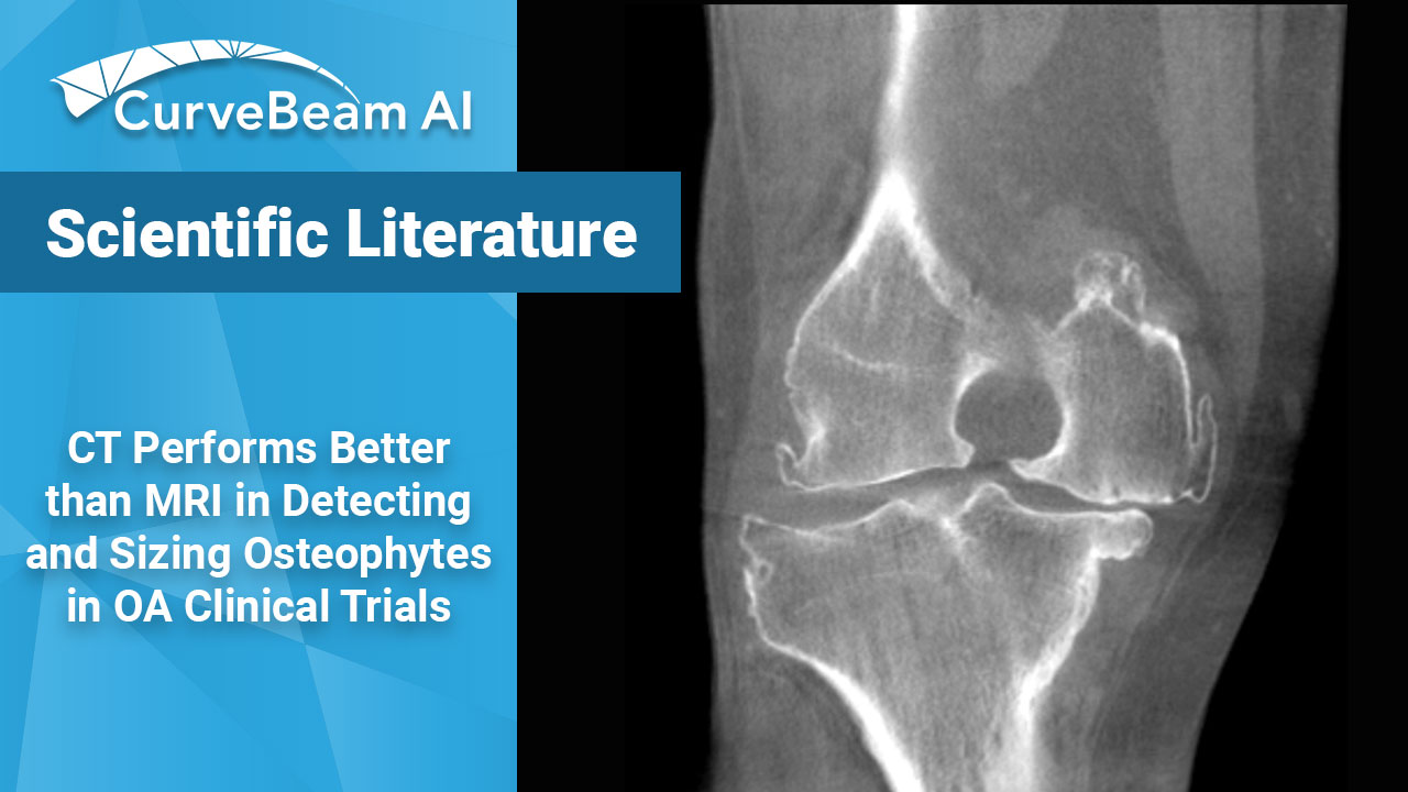
CT Outperforms MRI in Osteophyte Detection in Knees
Key Points:
- While magnetic resonance imaging (MRI) adequately detects the size and presence of osteophytes (OPs) in the medial tibio-femoral compartment, it underestimates OPs in all other knee compartments of osteoarthritic patients.
- CT may be helpful in the assessment of small osteophytes in all parts of the knee, particularly in early disease stages of OA.
Osteophytes (OP) are bony lumps that form in the knee bones and joints and are used to diagnose knee osteoarthritis (OA) on X-Rays.
Clinical trials will often use MRI to assess and grade OPs in arthritic knees. However, because the modality weights anatomical features based on density, OPs may be confused for fibrous or fatty soft tissue.
Computed Tomography (CT) is considered the imaging method of choice for analyzing mineralized structures such as OPs in all three knee compartments.
Dr. Roemer et al. out of the University of Erlangen-Nuremberg, Erlangen, Germany, conducted a study to rate the diagnostic performance of routine MRI in observational studies and clinical trials of knee OA against CT. They found that MRI not only underestimates the presence of OPs, but also the size of them, in all three knee compartments.
The researchers used clinical data from the Strontium Ranelate Efficacy in Knee Osteoarthritis (SEKOIA) trial to make the comparison. OPs were scored using a modified MRI Osteoarthritis Knee Score (MOAKS) in the patellofemoral (PFJ), the medial tibio-femoral (TFJ), and the lateral TFJ. OP Size was assessed in 18 locations in the knee.
Results showed that across 1098 locations evaluated, the PFJ MRI only detected 69% of OPs that CT defined, medial TFJ MRI detected 77% OPs that CT defined, and the lateral compartment MRI only detected 68% of OPs that CT defined.
When it came to OP size, the PFJ MRI underestimated the size 21% of the time. The medial TFJ MRI underestimated the size of the OPs 20% of the time. The lateral TFJ MRI underestimated OP size 15% of the time.
Regarding diagnostic performance for any OP detection, the medial TFJ MRI had a sensitivity of 0.91 and a specificity of 0.67.
Researchers concluded that MRI does in fact underestimate the presence and size of OPs in all 3 knee compartments. The medial compartment is considered the most relevant in disease-modifying OA drug trials; and MRI did demonstrate a sensitivity of >0.9 in that region. It was noted, however, that CT can be particularly helpful when assessing small OPs during early onset of the disease.
In the research setting, weight bearing CT (WBCT) imaging could be a useful tool. WBCT requires less infrastructure modifications than conventional CT and is simple to operate. Because WBCT uses a low power X-Ray source, patients are exposed to less dose than they would be in a conventional CT exam. As a bonus, researchers can observe joint space narrowing across the knee joint.
CurveBeam AI is the industry leader in WBCT imaging. CurveBeam AI’s HiRise provides functional 3D imaging of the entire lower extremities.
Click here to read about a new proposed OA classification method using WBCT.
Roemer FW, Engelke K, Li L, Laredo JD, Guermazi A. MRI underestimates presence and size of knee osteophytes using CT as a reference standard. Osteoarthritis Cartilage. 2023 Feb 14:S1063-4584(23)00594-0. doi: 10.1016/j.joca.2023.01.575. Epub ahead of print. PMID: 36796577.
Conti, Matthew S. MD; Ellis, Scott J. MD. Weight-bearing CT Scans in Foot and Ankle Surgery. Journal of the American Academy of Orthopaedic Surgeons 28(14):p e595-e603, July 15, 2020. | DOI: 10.5435/JAAOS-D-19-00700
Related Posts


