It is feasible for a single-practitioner podiatry practice to add weight bearing CT (WBCT) imaging and realize economical…
WBCT Indications Series: Lisfranc
Lisfranc Injuries
A Lisfranc injury is characterized by an injury to the ligament that connects the medial cuneiform with the 2nd metatarsal base (Lisfranc ligament). Often, there are associated midfoot fractures. Unstable Lisfranc injuries left untreated can result in chronic pain and progressive collapse of the midfoot.
A weight bearing CT scan can:
- Better characterize bony injuries1.
- Evaluate the 3D Lisfranc joint complex under physiologic load.
- Identify subtle Lisfranc injuries by effectively differentiating between stable and unstable Lisfranc injuries2.
Diagnosis
The diagnosis of Lisfranc injuries may be challenging on plain radiographs alone. A bilateral WBCT scan can be used to compare the injured to the uninjured side to detect subtle widening between the medial cuneiform and 2nd metatarsal base. A WBCT scan may also provide additional information about concomitant injuries and fractures.
Treatment Planning
WBCT can help determine if the injury pattern is more amenable to open reduction internal fixation (ORIF) or arthrodesis.
It can also be utilized to evaluate the 1st, 2nd, and 3rd tarsometatarsal (TMT), intertarsal, and intermetatarsal joints for instability and fractures.
Postoperative Assessment
For postoperative assessment of operative treatment of Lisfranc ligament injuries, a WBCT can:
- Accurately assess adequacy of reduction3.
- Accurately assess for any residual midfoot instability or persistent gapping between the medial cuneiform and 2nd metatarsal base4.
- Accurately assess healing of TMT joint fusions5.
Subtle Lisfranc Ligament Injury
40 yo female with low energy foot injury who presents with midfoot pain. Radiographs were indeterminate.
WBCT scan demonstrated widening between the medial cuneiform and second metatarsal base (red line).
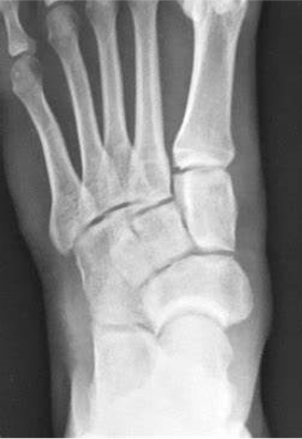
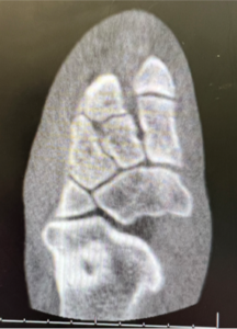
There were no fractures identified on the WBCT. She underwent ORIF of her Lisfranc injury.
Post-Op Assessment
34 yo male who suffered a motor vehicle accident four years ago presented with midfoot pain. He underwent midfoot fusion to address his instability.
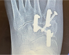
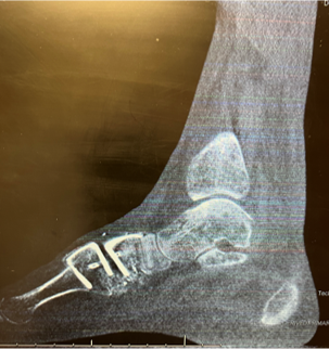
A postoperative WBCT scan demonstrated adequate reduction and fusion of the naviculocuneiform, 1st TMT, 2nd TMT, and intercuneiform joints.
Click Here to get direct links to the latest studies using WBCT scans.

Dr. Kent Ellington, MD
Dr. Kent Ellington, an OrthoCarolina Foot and Ankle Surgeon, has more than two decades of research experience in his field and is a well-respected name in diagnosis and treatment of below-the-knee conditions.
(1) Sripanich Y, Weinberg M, Krähenbühl N, Rungprai C, Saltzman CL, Barg A. Change in the First Cuneiform-Second Metatarsal Distance After Simulated Ligamentous Lisfranc Injury Evaluated by Weightbearing CT Scans. Foot Ankle Int. 2020 Nov;41(11):1432-1441. doi: 10.1177/1071100720938331. Epub 2020 Aug 20. PMID: 32819160.
(2) Lange, B., & Voldby, H. (2022, February 24). Webinar recap: WBCT scans of potentially unstable. CurveBeam AI. Retrieved March 30, 2023, from https://curvebeamai.com/webinars/webinar-recap-wbct-scans-of-potentially-unstable-weberbser2-fractures/
(3) Business Insider. (2021, May 20). MedShape and Curvebeam announce initiation of joint prospective clinical study. Business Insider. Retrieved March 30, 2023, from https://markets.businessinsider.com/news/stocks/medshape-and-curvebeam-announce-initiation-of-joint-prospective-clinical-study-1030408770
(4) Bhimani R, Sornsakrin P, Ashkani-Esfahani S, Lubberts B, Guss D, De Cesar Netto C, Waryasz GR, Kerkhoffs GMMJ, DiGiovanni CW. Using area and volume measurement via weightbearing CT to detect Lisfranc instability. J Orthop Res. 2021 Nov;39(11):2497-2505. doi: 10.1002/jor.24970. Epub 2021 Jan 6. PMID: 33368556.
(5) Steadman J, Sripanich Y, Rungprai C, Mills MK, Saltzman CL, Barg A. Comparative assessment of midfoot osteoarthritis diagnostic sensitivity using weightbearing computed tomography vs weightbearing plain radiography. Eur J Radiol. 2021 Jan;134:109419. doi: 10.1016/j.ejrad.2020.109419. Epub 2020 Nov 21. PMID: 33259992.


