
AI-Driven Segmentation on Weight Bearing CT Introduction Accurate segmentation is critical for orthopedic workflows, including…
Bernasconi & Lintz: WBCT Should be First-Line Imaging for Cavovarus Deformity
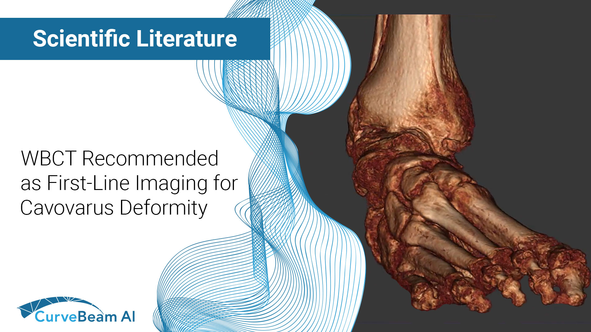
Cavovarus deformity in the foot involves the forefoot, midfoot, hindfoot, and often the talus. Because the deformity is so complex, weight bearing CT can be a tremendous tool to evaluate patients with this condition.
In a recent paper published in Foot and Ankle Clinics, authors Alessio Bernasconi and Francois Lintz even suggest weight bearing CT should be the first-line imaging modality for cavovarus patients because of the advantages it offers for surgical planning.
In cavovarus patients, the bean shape of the deformity means there is severe bone superimposition and abnormal joint rotation. Add in technologist bias and acquiring adequate weight bearing X-Rays can be challenging. While conventional medical CT helps with evaluating joint status, it exposes patients to higher radiation and provides no alignment information.
With weight bearing CT, we can measure cavovarus malalignment with either classic measurements made on single slices or with automated 3D measurements. 3D measurements could help us better understand the whole deformity, especially the center of rotation and angulation, or CORA, of the joints. This is especially important as Cavovarus could be thought of as a multi-CORA condition, according to the authors.
Current treatment for cavovarus foot generally includes calcaneal osteotomies and dorsiflexion osteotomies, but these approaches aim to correct global mechanical balance rather than the CORA. This may explain why some of these surgeries fail. Weight bearing CT imaging could be key to shifting to a surgical approach to target the true CORA, Drs. Bernasconi and Lintz wrote.
As for evaluating joint status, distance mapping tools for weight bearing CT measure the distance distribution between articular surfaces of the joints. Distance mapping in daily practice could provide a much more objective assessment of joints than the Kellgren Lawrence grade, and could help surgeons identify specific overloaded areas when planning realignment procedures, the authors wrote.
What new insights will weight bearing CT provide in foot, knee and hip deformities? We’ll keep an eye on the research and keep you updated.
Access the full study here: https://pubmed.ncbi.nlm.nih.gov/36585514/
Related Posts


