At CurveBeam AI, we often talk about the clinical and workflow advantages of weight-bearing CT,…
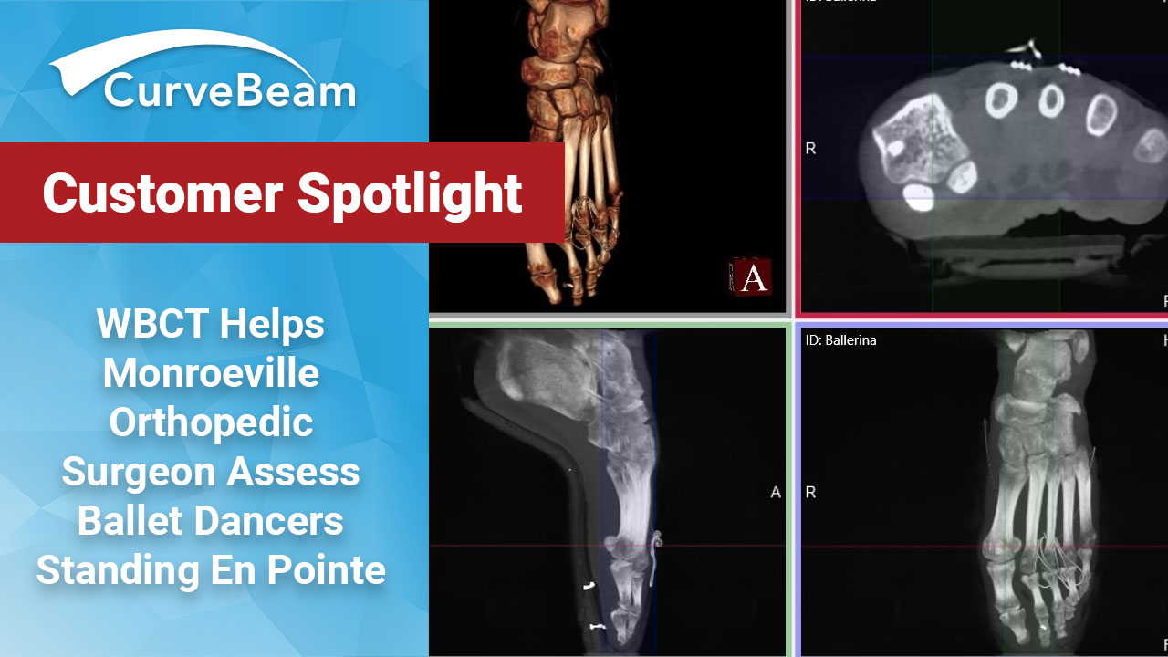
Weight Bearing CT Helps Monroeville Orthopedic Surgeon Assess Ballet Dancers Standing En Pointe
As a soloist for the Pittsburgh Ballet Theater, dancer Diana Yohe, 25, has sprained her ankle several times – enough times that she had to have a ligament reconstruction surgery on her left ankle.
Throughout her recovery, her surgeon ordered X-Ray exams to track the progress of her healing. Yet Yohe worried she might not be ready for her upcoming lead role in Giselle.

Shortly before opening night, Dr. Victor Prisk, MD, ordered a state-of-the-art standing CT exam. Yohe stood en pointe during the scan.
“I could literally see the bones, the ligaments and even the skin,” Yohe said. “Prior to surgery my ankle was badly skewed. But on the CT scan, I could see it was back to perfect alignment.”
 Dr. Prisk specializes in foot & ankle sports and dance medicine. He recently added weight bearing CT technology to Prisk Orthopedics and Wellness, PC, his private practice in Monroeville, PA. During a weight bearing CT scan, a patient stands naturally while the X-Ray tube and detector rotates 360-degrees around their feet & ankles. The X-Rays are then processed to produce a three-dimensional image of the patient’s foot.
CT imaging is more sensitive than 2D X-Rays in detecting fractures, fracture healing, and other conditions. However, traditional CT technology typically requires much more infrastructure than the average orthopedic clinic can accommodate and exposes the patient to a higher radiation dose than 2D X-Ray. In addition, foot & ankle specialists require a standing or “weight bearing” view of the foot and ankle for most conditions. In traditional CT imaging, the patient is lying down.
Dr. Prisk’s weight bearing CT scanner fits in his clinic’s X-ray room, and peer reviewed research demonstrates the scanner’s radiation dose is comparable to an X-Ray exam.
“I invested in this technology because I knew it would be a game changer in how I look at the foot & ankle from a 3D perspective; it literally adds another dimension,” Dr. Prisk said. “It allows me to critically assess deformities like never before.”
Dr. Prisk offers a pedCAT exam for every patient during their initial visit.
“The pedCAT allows me to get all X-Ray views for a new patient in less than 45 seconds, and is higher quality than standard X-Ray imaging,” Dr. Prisk said.
Several of Prisk’s patients are ballet dancers. With the pedCAT, he can get a 3D X-Ray while the dancer is standing en pointe.
“Scanning a foot en pointe allows to me see mal-alignments that could be resulting from an intrinsic foot problem or even from ballet technique,” Prisk said. He added there have been cases where he was able to determine the ballet dancer was wearing ill-fitting pointe shoes, as the toe box was either too large or too small.
Dr. Prisk, a former professional dancer himself, sprained his ankle during a gymnastics practice when he was 15 years old. The injury inspired him to become an orthopedic surgeon.
“My dance career taught me how prevalent foot & ankle injuries were for these athletes, and I wanted to help this particular population,” Dr. Prisk said. “Perhaps back then, if pedCAT technology were available, I would have known if my own ankle was displaced.”
Yohe is back to dancing five days a week. She said her first performance of Giselle went really well.
Related Posts