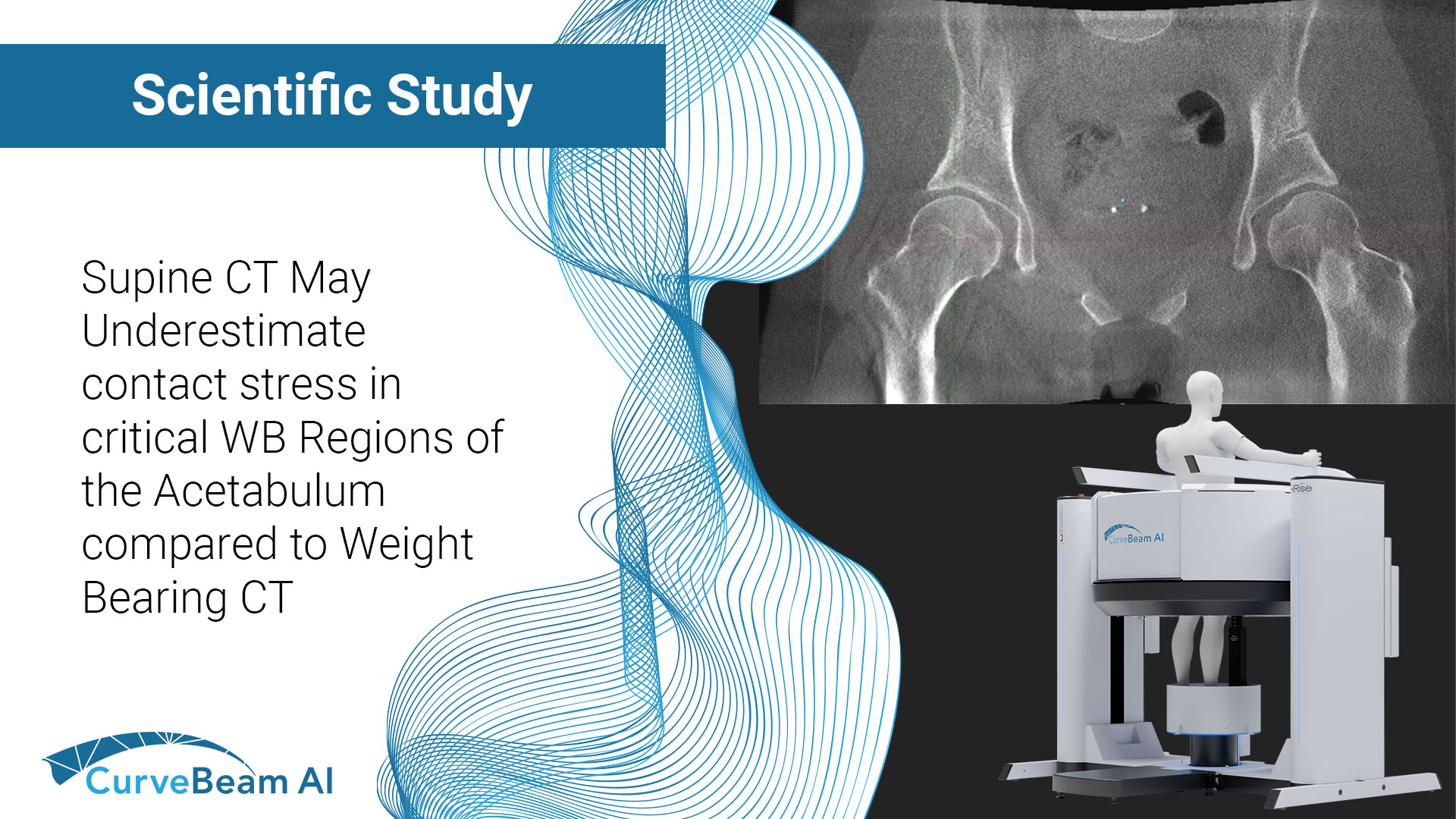
Supine CT May Underestimate Contact Stress in Critical WB Regions of the Acetabulum Compared to Weight Bearing CT
Key Points: Computational models of the hip often omit patient-specific functional orientation when placing imaging-derived bony geometry into anatomic landmark-based coordinate systems for application of joint loading schemes. Significant differences are found when incorporating WBCT-derived data suggesting non-weight bearing (NWB)…
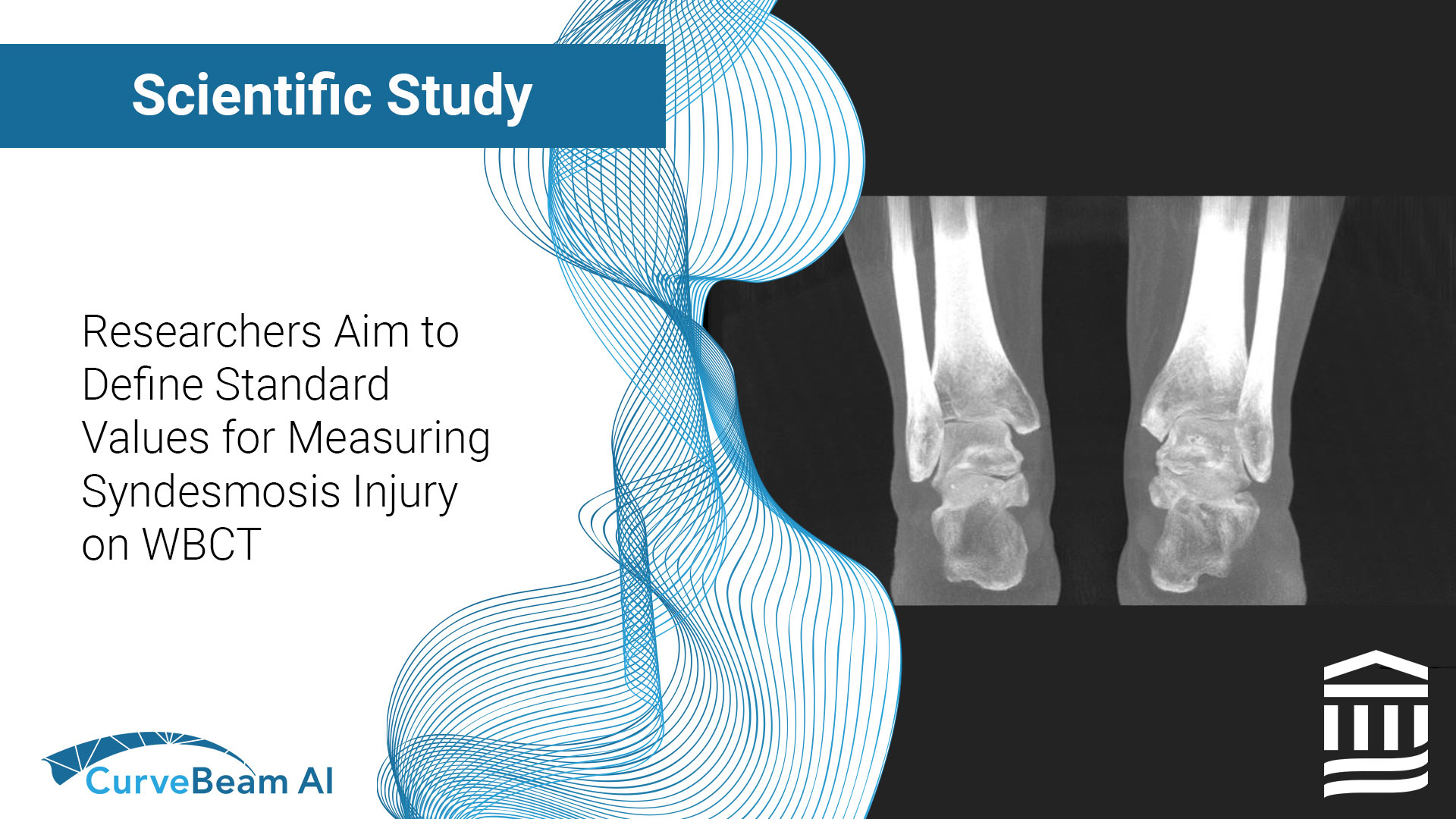
Researchers Aim to Define Standard Values for Measuring Syndesmosis Injury on WBCT
Key Points: Weight Bearing CT (WBCT) allows for bilateral comparative data that is imperative when diagnosing ankle syndesmosis. Side-to-side volume differences of 19% or greater may be indicative of an abnormality. Gender norms are important to consider when evaluating ankle…
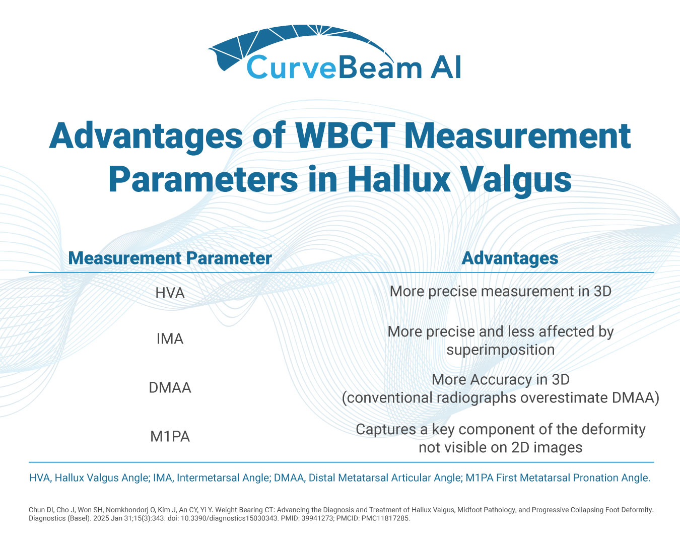
Advantages of WBCT Measurement Parameters in Hallux Valgus
When it comes to hallux valgus (bunion) deformity, weight bearing CT provides significant advantages over conventional radiography. "Its ability to provide detailed 3D visualization, coupled with automated measurement techniques and AI integration, allows for a more precise assessment of the…
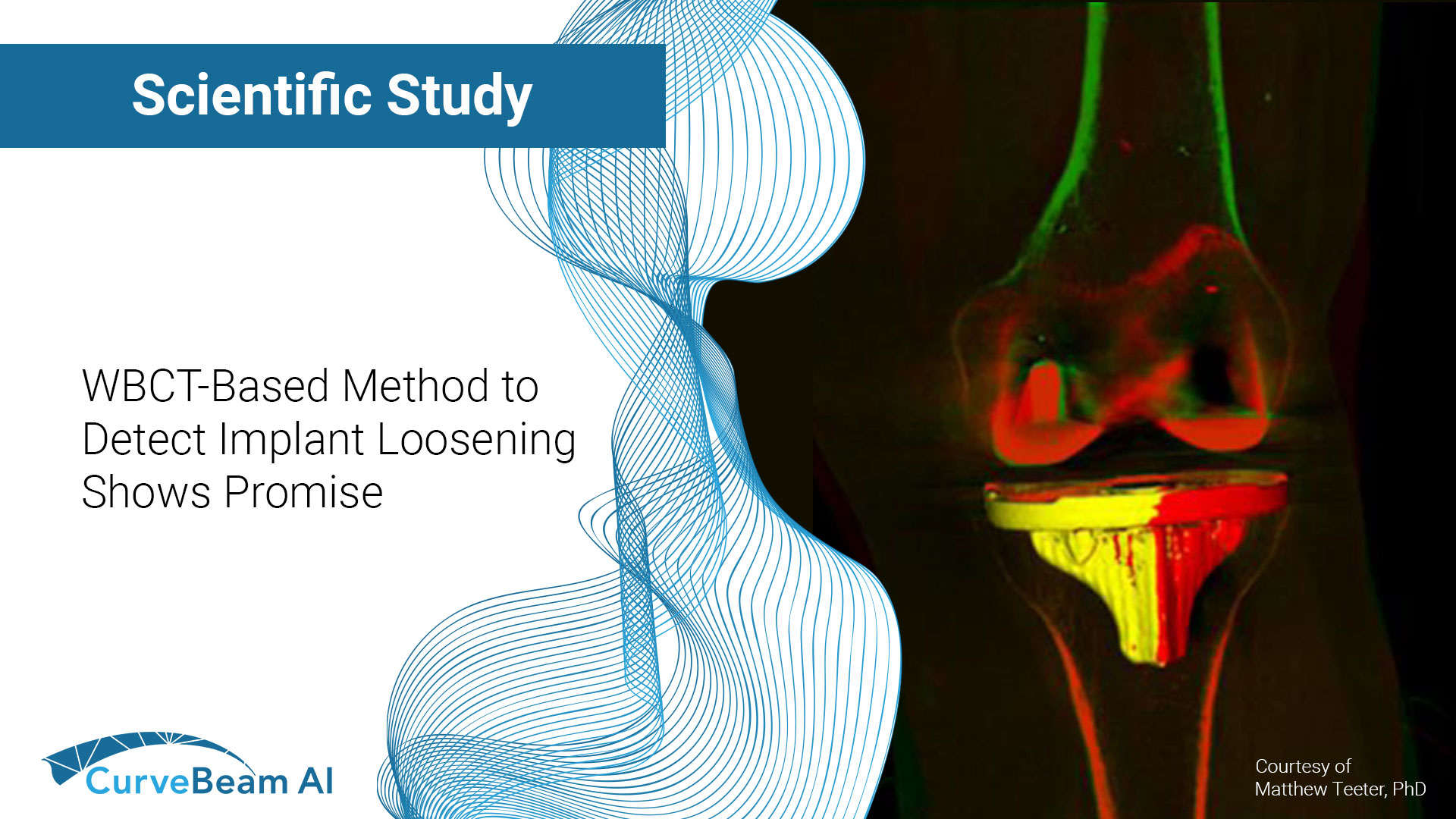
WBCT-Based Method to Detect Implant Loosening Shows Promise
Key Points: Radiostereometric Analysis (RSA) uses X-Ray and markers inserted into the bone intraoperatively to assess implant migration and fixation. Model-based RSA (MBRSA) improves upon RSA by incorporating 3D implant CAD models. In one study, weight bearing CT (WBCT)-RSA was…
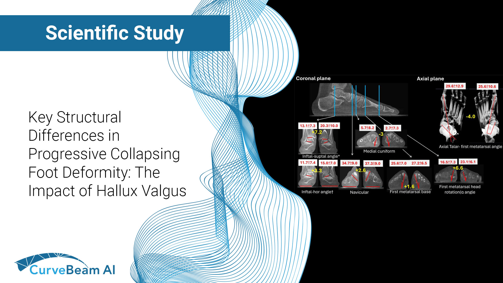
Comparative Analysis of Structural Differences in Progressive Collapsing Foot Deformity With and Without Hallux Valgus
Key Points: Weight Bearing CT (WBCT) helps visualize several structural and alignment differences between Progressive Collapsing Foot Deformity (PCFD) with and without Hallux Valgus (HV). Rotation of the first tarsometatarsal joint, and intrinsic rotation of the first metatarsal bone were…
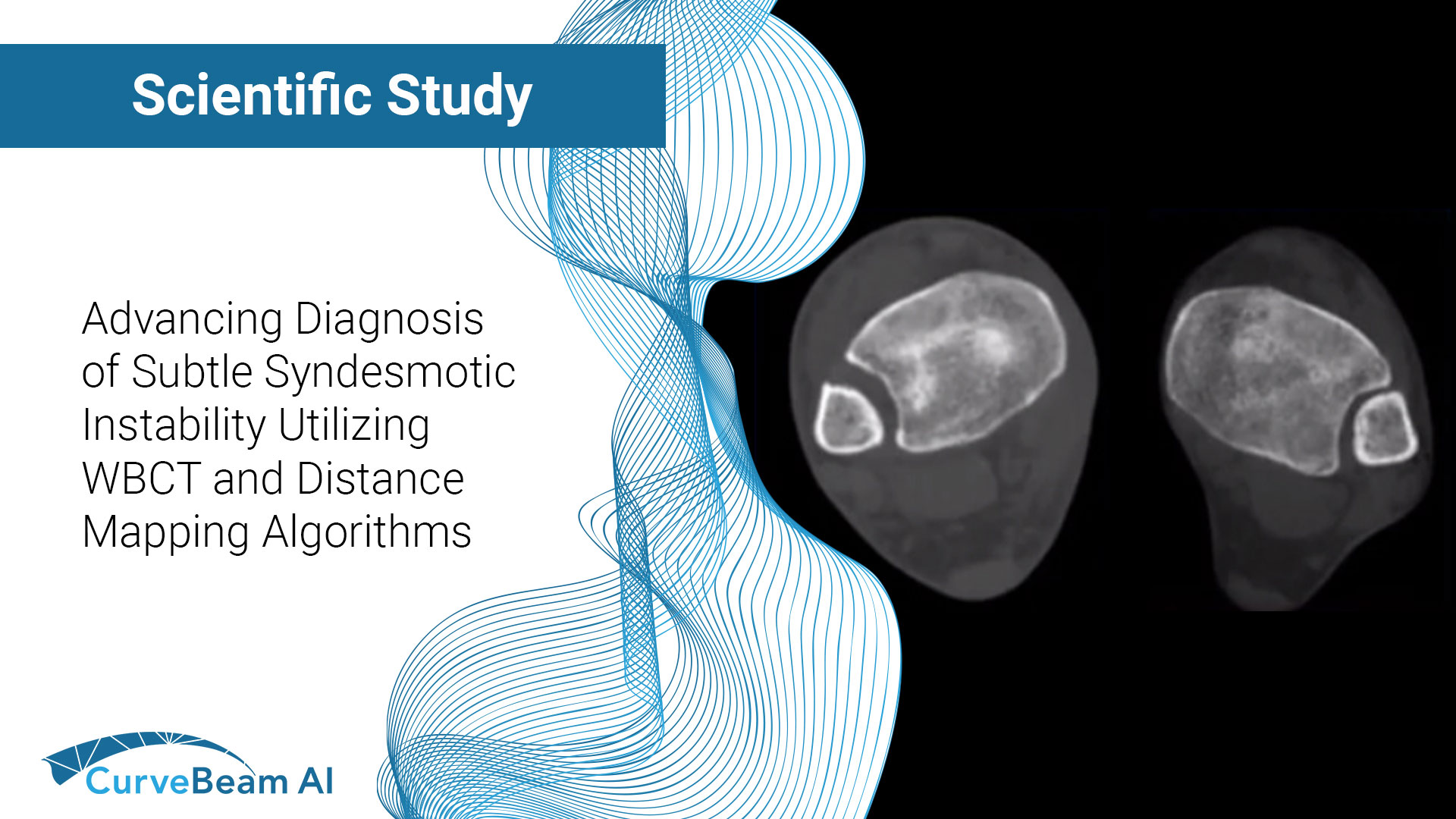
Cadaveric Diagnostic Study of Subtle Syndesmotic Instability Using a 3-Dimensional Weight Bearing CT Distance Mapping Algorithm
Key Points: Imaging modalities such as plain radiographs (X-Ray), computed tomography (CT), and magnetic resonance imaging (MRI), don’t have the diagnostic accuracy needed to detect syndesmotic widening or subtle instability. Weight Bearing CT (WBCT) imaging when combined with a distance…
Congruent Weber-B Ankle Fractures Do Not Alter Tibiotalar Contact Mechanics
Key Points: Weight Bearing CT (WBCT) provides critical data otherwise missed in weight bearing radiographs when trying to understand the true impact of a fracture on joint stability. Computational biomechanics, when paired with bilateral WBCT imaging, provides a better understanding…
Cone Beam Weight Bearing Computed Tomography of Ankle Arthritis and Total Ankle Arthroplasty
Key Points: Cone Beam CT (CBCT), specifically WBCT three-dimensional (3D) visualization approach allows for a more accurate assessment of foot alignment with attention to planus, cavus, varus or valgus deformities. Post-operatively, weight bearing CT (WBCT) has the potential to better…
Estimation of Radiographic Joint Space of the Trapeziometacarpal Joint with Computed Tomographic Validation
Key Points: Cone Beam Computed Tomography (CBCT), a three-dimensional (3D) imaging modality, can readily evaluate complex joint structures. The most important radiographic (X-Ray) criterion used in surgical decision making for osteoarthritis is joint space narrowing (JSN), or the loss of…