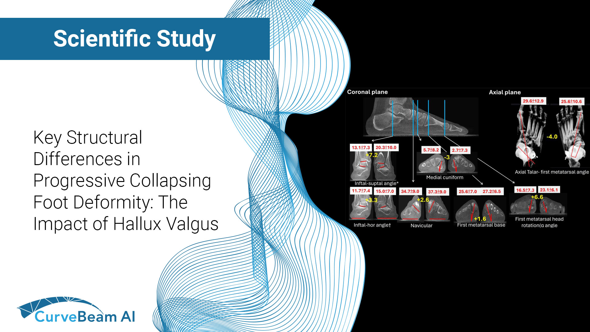
Transforming Bunion Assessment with Weight Bearing CT
By Dr. David Soomekh, DPM Foot and ankle specialist and surgeon, sports medicine & reconstructive foot and ankle surgery, Foot & Ankle Specialty Group, Beverly Hills, CA
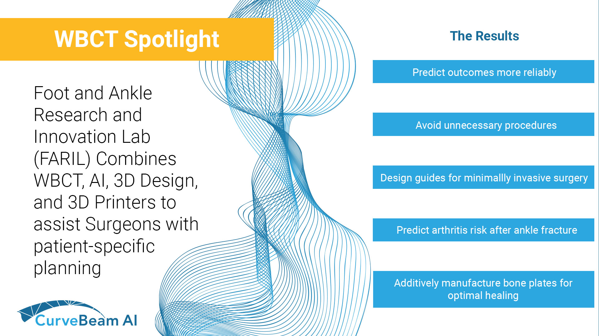
Key Points: Cone Beam CT (CBCT), specifically WBCT three-dimensional (3D) visualization approach allows for a more accurate assessment of foot alignment with attention to planus, cavus, varus or valgus deformities. Post-operatively, weight bearing CT (WBCT) has the potential to better…
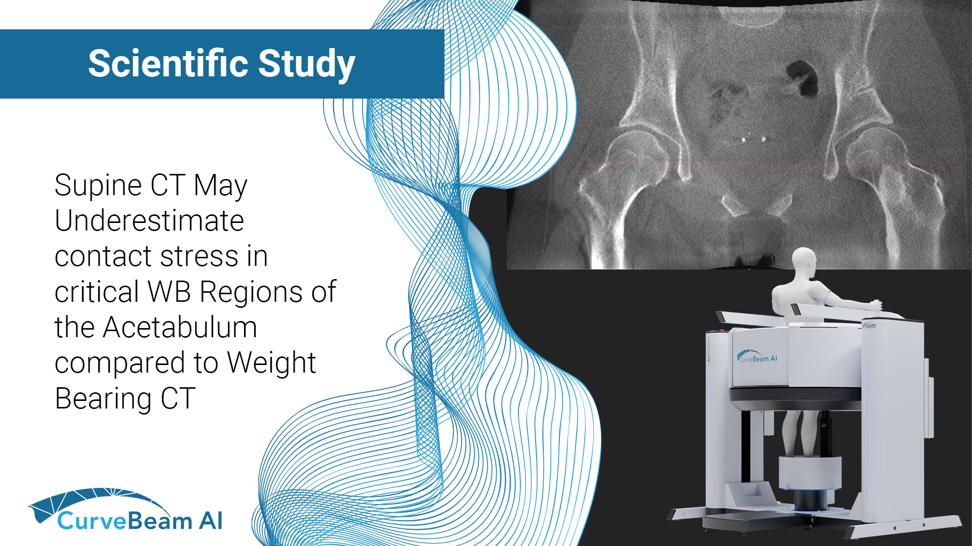
The HiRise is a low dose cone beam CT system. The upper extremity scans are quick and allow for patients to be scanned in a comfortable seated position.
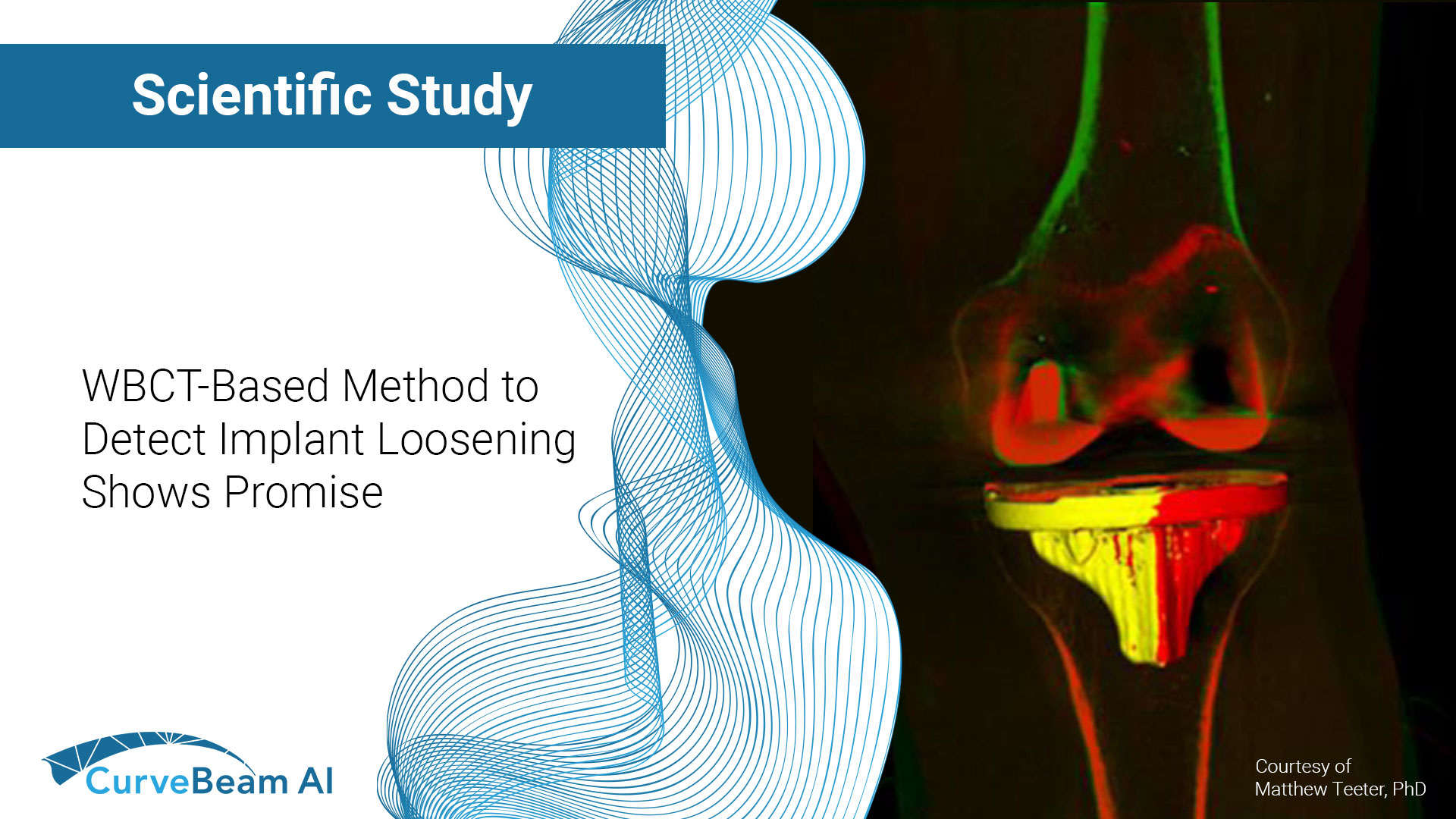
Key Points: Cone Beam Computed Tomography (CBCT), a three-dimensional (3D) imaging modality, can readily evaluate complex joint structures. The most important radiographic (X-Ray) criterion used in surgical decision making for osteoarthritis is joint space narrowing (JSN), or the loss of…
Key Points: Cone Beam CT (CBCT) is superior in assessing bony structures compared to magnetic resonance imaging (MRI) In this study, there was a 40% rate of discrepancy when grading knee subchondral insufficiency fractures on CBCT vs. MRI, with MRI…
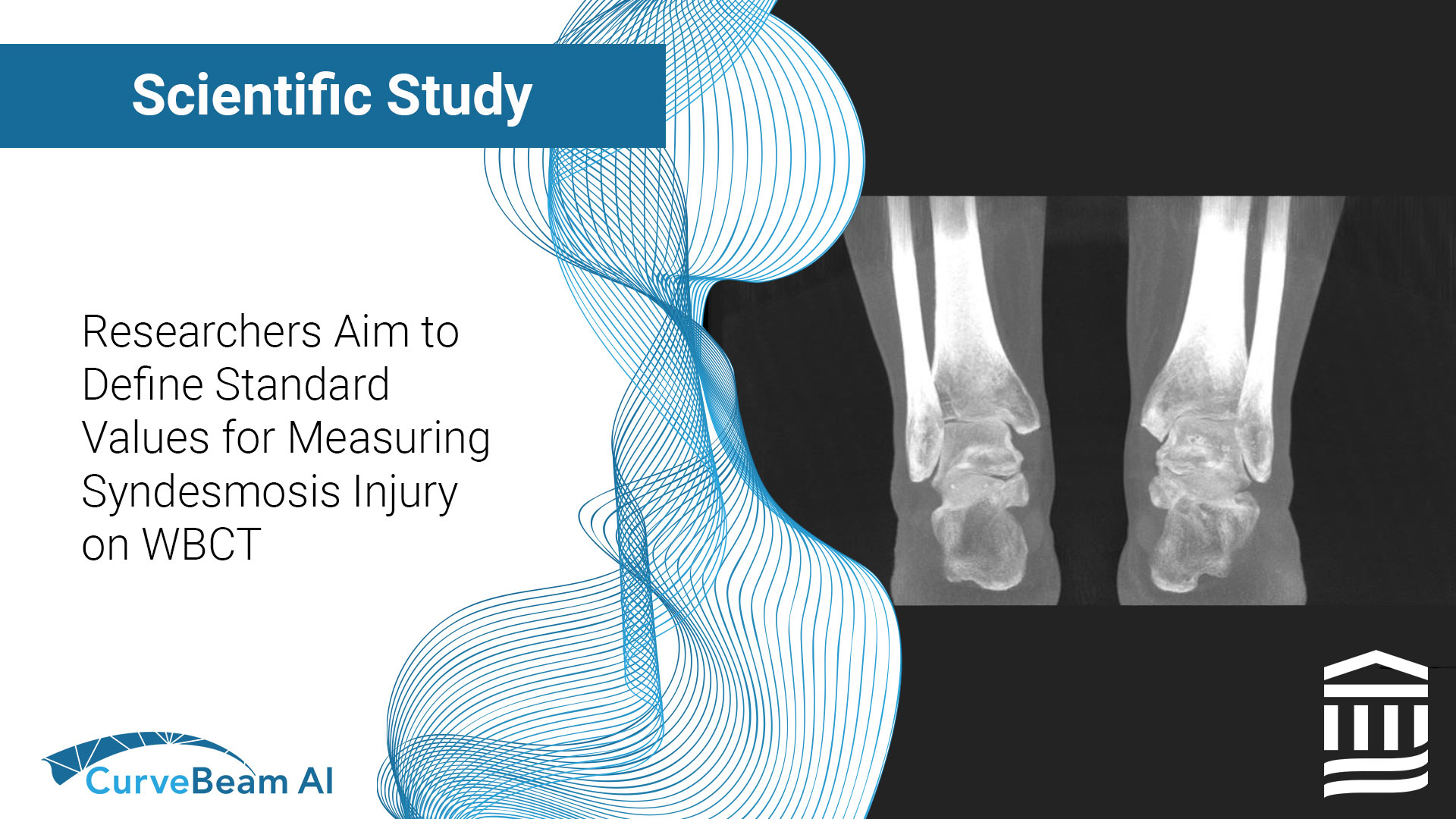
Key Points: WBCT 3D Measurements volumetric measurements of the syndesmosis have shown to have a high specificity (83.3%) and sensitivity (95.8%) for the diagnosis of subtle injuries. Manual 3D WBCT volume measurement of syndesmosis, a promising method to detect instability,…