Orthopedic decision-making depends on accurate representation of anatomy—particularly when joint alignment and bone relationships change…

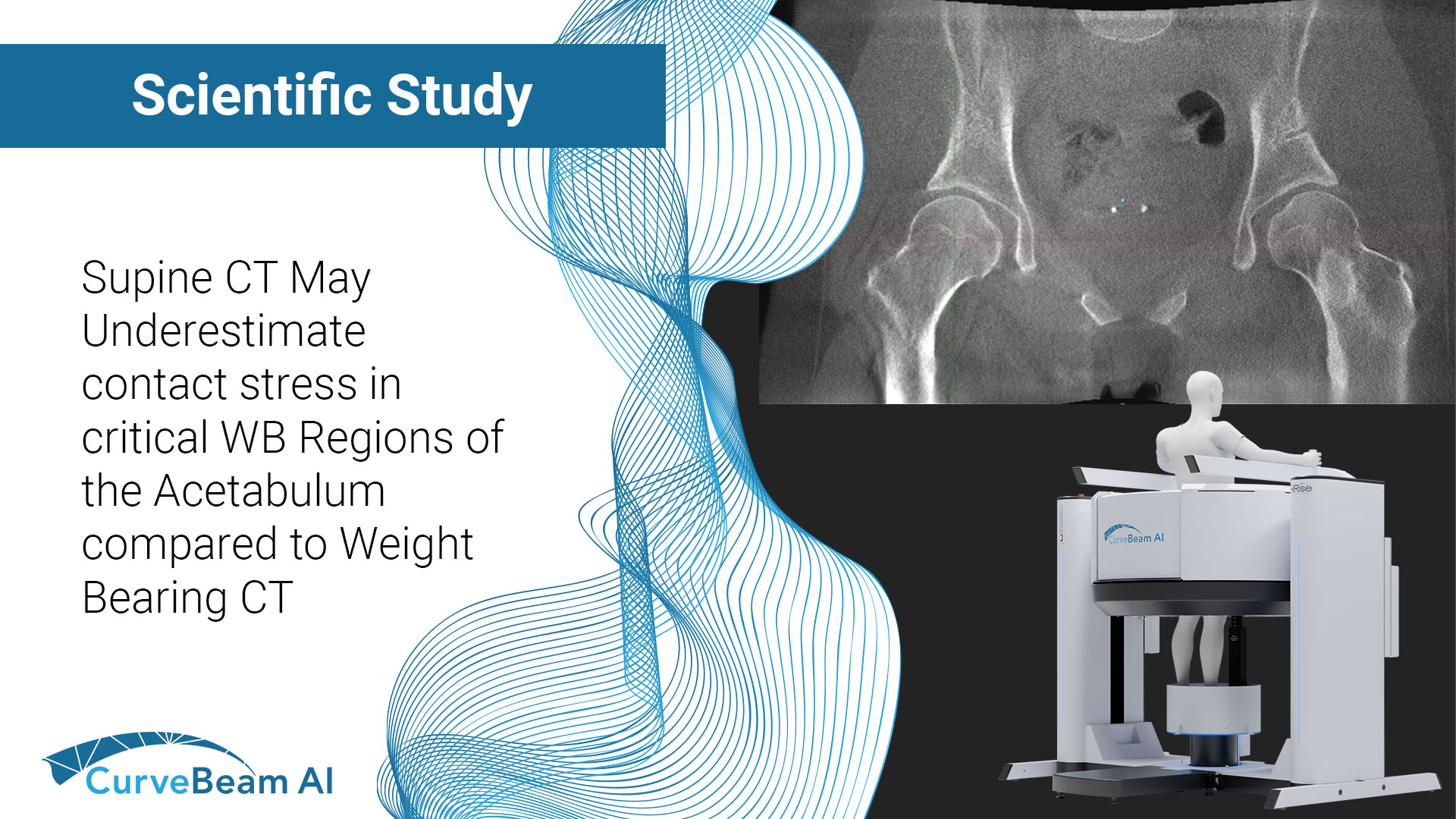
Supine CT May Underestimate Contact Stress in Critical WB Regions of the Acetabulum Compared to Weight Bearing CT
Key Points:
- Computational models of the hip often omit patient-specific functional orientation when placing imaging-derived bony geometry into anatomic landmark-based coordinate systems for application of joint loading schemes.
- Significant differences are found when incorporating WBCT-derived data suggesting non-weight bearing (NWB) methods could result in underestimating contact stress in critical weight bearing regions of the acetabulum.
- WBCT presents the opportunity to increase patient specificity and thus accuracy of computed contact mechanics without the need to perform adjunctive procedures such as full kinematic and kinetic gait analysis.
Hip dysplasia is a musculoskeletal condition that results in insufficient coverage of the femoral head by the acetabulum. This lack of coverage leads to hip pain and accelerated progression of osteoarthritis. It is presumed that this happens due to instability and abnormalities in the force distribution within the hip joint.
Computational modeling techniques such as finite element analysis (FEA) and discrete element analysis (DEA) are commonly used to evaluate variations in cartilage contact stress that result from abnormal force distribution in a variety of joints. These methods have shown to provide reliable and accurate contact stress calculations in cadaveric validation studies performed in the hip.
Computational models of living patients are typically generated from clinical NWB supine CT or MRI scans, which require scans to be reoriented to a neutral, standardized pose before functional load application. The reorientation process involves translating the 3D models to a standardized coordinate system of bone landmarks. This standardization removes critical elements such as patient-specific femoral version and pelvic tilt during weight bearing from the resulting computational models.
Methods such as two-dimensional (2D) weight bearing radiographs (X-Ray) have been able to incorporate standing pelvic tilt into patient-specific FEA models resulting in computation of decreased hip contact area and increased median contact pressure. However, even with this technique, other potentially relevant features of an individual’s acetabular-femoral relationship (hip extension/abduction) are omitted.
Similar to weight bearing X-Rays, WBCT captures functional joint position in 3D while a patient is standing in a natural weight bearing position. Beyond providing relatively simplistic measurements of joint alignment,
WBCT holds the exciting potential for providing the data to generate more accurate patient-specific computational models of joints.
This is the first study to create computational models of the hip. Dr. Dominic J.L. Rivas et al, out of the Department of Orthopedics and Rehabilitation at the University of Iowa, in Iowa City, Iowa, USA set out to evaluate how joint contact mechanics computed from models based on an upright, WBCT-derived, patient-specific orientation differed from mechanics computed from models developed by placing clinical CT-derived geometry into a standardized, anatomic landmark-based orientation. Researchers hypothesized that inclusion of a patient-specific weight bearing pose would result in calculation of higher contact stress and differences in contact area in different locations within the acetabulum.
Methods
Ten hip dysplasia patients indicated for periacetabular osteotomy (11 hips) consented to a preoperative, research-specific pelvic WBCT scan on the HiRise system. Standard clinical supine CTs for the same patients were obtained from the patients’ medical record. The pelvis and femur from each patient’s non-WBCT scan was aligned to the International Society of Biomechanics (ISB) coordinate system (NWB-ISB). The original NWB geometry was also registered to the WBCT scan. Three additional WBBCT-dervied patient specific models were generated for each hip to account for realistic axial and coronal rotation in gait loading. Computed contact stress and contact area were compared between model initialization techniques.
Results
It was found that the addition of sagittal tilt did not significantly change whole-joint peak or mean contact stress or contact area. Inclusion of motion-captured coronal and axial rotation decreased peak contact stress and slightly increased average contact area from WBCT-sagittal models. Including all WBCT-derived rotations further reduced computed peak contact stress and significantly increased contact area. Variably significant differences in patient-specific acetabular subregion mechanics indicated the importance of functional orientation incorporation for modeling applications in which local contact mechanics are of interest.
Conclusion
Researchers concluded that incorporation of WBCT-derived patient-specific stance into computational models of dysplastic hips produced modest differences in computed whole-joint contact stress, which suggests that the approach of using anatomic landmark-based coordinate systems is acceptable when considering peak or mean contact stress on a whole-joint scale.
Total contact area differed significantly between model initiation techniques, and inclusion of patient-specific pelvis/femur orientation from the WBCT caused variable, patient-by-patient, regional effects on the computed contact area and stress metrics. These variable mechanical differences introduced the potential to underestimate contact stress in critical weight bearing regions of the acetabulum within the context of an overall reasonable approximation of whole-joint contact stress. Such localized differences could have substantial implications for focal cartilage degeneration.
It was noted that WBCT presents the opportunity to increase patient specificity and thus accuracy of computed contact mechanics without the need to perform adjunctive procedures such as full kinematic and kinetic gait analysis.
To read the full study click here.


