Chat with us, powered by LiveChat Skip to content

Case Study: pedCAT vs. Radiographs

A patient sought a second opinion for the cause of her medial foot and ankle pain. The treating physician used plain radiographs to diagnose her with posterior tibial tendon dysfunction/partial tear and degenerative joint disease of the 1st, 2nd, and 3rd tarsometatarsal joints. The treating physician also noted a “chip” of bone on the inside of the ankle. The treating physician planned on performing a flat foot reconstruction, a posterior tibial tendon repair, and a tarsometatarsal joint fusion. The physician performing the second opinion noted the patient’s discomfort over the medial ankle gutter was much more significant than over the posterior tibial tendon and the spring ligament. The patient had minimal discomfort through the tarsometatarsal joints. The physician performing the second opinion ordered a weight bearing pedCAT study to assess the midfoot DJD and to better evaluate the midtarsal joint and ankle joint. The pedCAT study clearly documented a degenerative process in the medial ankle gutter with a bony impingement. On secondary exam, the majority of the symptoms arose from the medial ankle gutter. If the flat foot reconstruction was performed as planned, the talus would have been dorsiflexed and the tibio-talar impingement would have been worsened. The pedCAT images helped prevent an un-necessary surgery, while directing the physician to the appropriate pathology.
Read more

Distinguishing Categories of Medial Column Ligamentous Fracture

Weight bearing CT scans offer new, three-dimensional perspectives that reveal a better understanding of the biomechanics of flat foot. That was a finding on an academic poster on display at The American Orthotic and Prosthetic Association's National Assembly in Las Vegas (Sept. 4 - 9). "IRD, PTTD, Posterior Tib, Flat Foot, Pronation...these are all terms that various professions use to describe some sort of medial column ligamentous breakdown," according to the poster. "The purpose of this study is to better distinguish some types of pathomechanics of the foot. By categorizing and redefining them, it makes the description of pathology more useful for Orthotic Treatment. This was possible through the use of 3D rendering of the pedCAT machine and CurveBeam software." The study was conducted by Ian Engelman, M.S. CPO, and Harold Chamberlin, DPM. They looked at 3D renderings of the bony structures in flat feet, and came up with some new descriptions, as well as their possible ligamentous causes. The study concluded that while the exterior contours of the foot suggest certain bony pathologies, the 3D renderings provided by the pedCAT would allow for orthotists to better distinguish the pathomechanics of the foot and ankle. An unexpected result showed a moderately strong correlation (-0.68) between the vertical position of the navicular and medial displacement in a patient population with ranging degrees of medial column breakdown.
Read more

pedCAT: Weight Bearing CT Views of Lisfranc conditions

Lisfranc injuries can easily be missed on traditional radiographs. One research study tried to determine the level of accuracy when using plain radiographs to assess midfoot conditions. Their results confirmed a high incidence of missed diagnosis even by experienced observers.¹ “Radiographic evaluation is crucial in the diagnosis…Weight bearing views are more sensitive since tarso-metatarsal instability may be revealed…The threshold for cross sectional imaging such as multi-detector CT should be low,” according to the study. With the pedCAT, it is possible to get a weight bearing, three dimensional scan at the point of care. The tarso-metatarsal joint can be viewed without any superimposition from surrounding anatomy. Delaying a 3D scan can be detrimental to recovery. “The identification of Lisfranc joint injury can be difficult and is often not detected on initial presentation to the accident department. This is important because of the correlation between delay in treatment, particularly more than six months, and poor functional outcome,” the study reported. Click on the blog post title to see examples of weight bearing CT scans of Lisfranc injuries.
Read more

pedCAT: Fracture & Fusion Assessments

X-rays of the foot and ankle may not always provide conclusive assessments for post-operative fusions such as of the tarso-metatarsal joint or hind foot joints. Similarly, the physician is often left guessing if a fracture has properly healed. “The pedCAT takes all the variability out, all the guesswork out of it,” Dr. Martin O’Malley, MD, an Associate Attending Orthopedic Surgeon at the Hospital for Special Surgery in New York, New York, said. Dr. O’Malley said pedCAT scans allow him to clearly determine if a fusion has healed more than 50 percent, and he decides when to ambulate his patient accordingly. “I let people walk on it earlier than before, and I keep them off longer than before,” Dr. O’Malley said. The pedCAT provides a three-dimensional view of fractures that changes the way O’Malley sees this common diagnosis. “These posterior pieces are often bigger than we thought were based on plain X-ray and they often travel all the way around the medial side as well, which we never thought they did,” Dr. O’Malley said. “You know, we thought it was an infrequent fracture, but now we see it routinely. Now most of my ankle fracture work, I’d say more than half the time, is through a posterior approach. For the first 15 to 18 years of my practice I would do medial/ lateral incisions. Now I’m going to the back of the ankle. And a lot of it is driven by the pedCAT.” pedCAT Stress Fracture Cross Section Click on the blog post title to see an example of a navicular stress fracture in a collegiate runner that had not healed at all after six weeks of casting. Were it not for the conclusive pedCAT scan, this patient would have been allowed to ambulate. Watch Dr. O’Malley talk more about the pedCAT here . To offer your patients state-of-the-art fracture & fusion assessment, consider a pedCAT for your practice.
Read more
WorldCupSoccerPreview2

World Cup Special Edition: Soccer Player with a Sesamoid Injury

The 2014 FIFA World Cup officially begins Thursday, June 12. The world’s top soccer players will all be in Rio, Brazil for one of the biggest sporting events of the decade. The next two weeks will be filled with cheering fans, tears of joy, and national pride. But inevitably, there will also be injuries. In fact, before the games even begin, a number of players already know they will have to sit them out. For professional athletes and weekend warriors alike, an accurate diagnosis from the onset can make a huge impact on the speed of recovery. When it comes to bony injuries of the foot and ankle, the pedCAT is the only tool in the world that can provide a weight bearing, three dimensional image of the entire foot. Here’s a case of a 30-year-old soccer player, who was treated by a California podiatrist. The athlete injured his right great toe and sesamoids while playing soccer. The doctor observed that the patient’s first metatarsophalangeal joint was swollen and ecchymotic, and that he was tender upon palpation of the great toe joint and sesamoid bones. The doctor took standard non-weight bearing X-Ray images of the patient. The AP and lateral X-Rays showed an obvious fracture with displacement in the medial sesamoid. The lateral appeared to have a fracture, but the doctor could not confirm his suspicion with the X-Ray. The doctor also took an axial X-Ray, but it had no diagnostic benefit since the patient could not extend his great toe. double sesamoid fracture x-ray The doctor then took a pedCAT scan of the same patient. The CT images clearly showed a severely displaced right medial sesamoid oblique fracture and a minimally displaced lateral sesamoid fracture. The oblique lateral sesamoid fracture demonstrated 2 mm of plantar displacement with excellent bony contact/ apposition in the dorsal 4 mm of the sesamoid. pedCAT double sesamoid fracturepedCAT lateral sesamoid fracture displacement Although the diagnostic information provided by the pedCAT did not alter the treatment plan for the medial sesamoid, it did confirm the injury to the lateral sesamoid. More importantly, the pedCAT was able to provide diagnostic information regarding the geometric nature of the lateral sesamoid fracture. “I could determine that there was enough bony contact to allow for bone healing with appropriate conservative care management,” the doctor said.
Read more

pedCAT: Improved Outcomes

pedCAT: Improved Outcomes

The message at RSNA Annual Meeting this past November was clear: new US healthcare laws mean the practice of radiology is no longer about volume, but value.

“We are focusing on quality metrics. It’s becoming important for us to become champions of quality in our institutions,” said Dr. Vijay Rao, MD, in a course at the meeting.

How could a pedCAT add value to your practice?

We might get an idea by looking at a comparable new technology. Breast tomosynthesis mammography provides 3D imaging for breast cancer screening, similar to the way the pedCAT provides 3D imaging for the foot and ankle. Breast tomosynthesis technology can detect breast cancers earlier than traditional 2D mammography, and can more accurately pinpoint the size, shape and location of abnormalities, according to the Massachusetts General Hospital Imaging Department.

Dr. Liane Philpotts, professor of diagnostic radiology at the Yale School of Medicine, called tomosynthesis a “game changer” and a “win-win.”

In the same way, the pedCAT eliminates variability, helps lead to better diagnoses, and makes both you and your patients more confident that treatment will result in better outcomes.

Dr. Erik Nilssen, MD, said the pedCAT helps him determine exactly when to allow patients to ambulate, “based on our ability to monitor fracture healing and fusion rates.”

Also, pedCAT scans can take the guesswork out of hindfoot alignment, said Dr. Martin O’Malley, MD, because they allow for reproducible measurements.

“We’ve never had a reproducible measurement,” until now, he said.

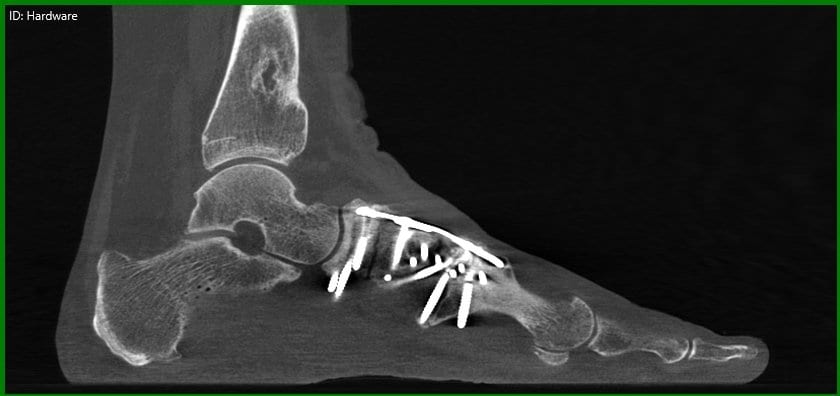
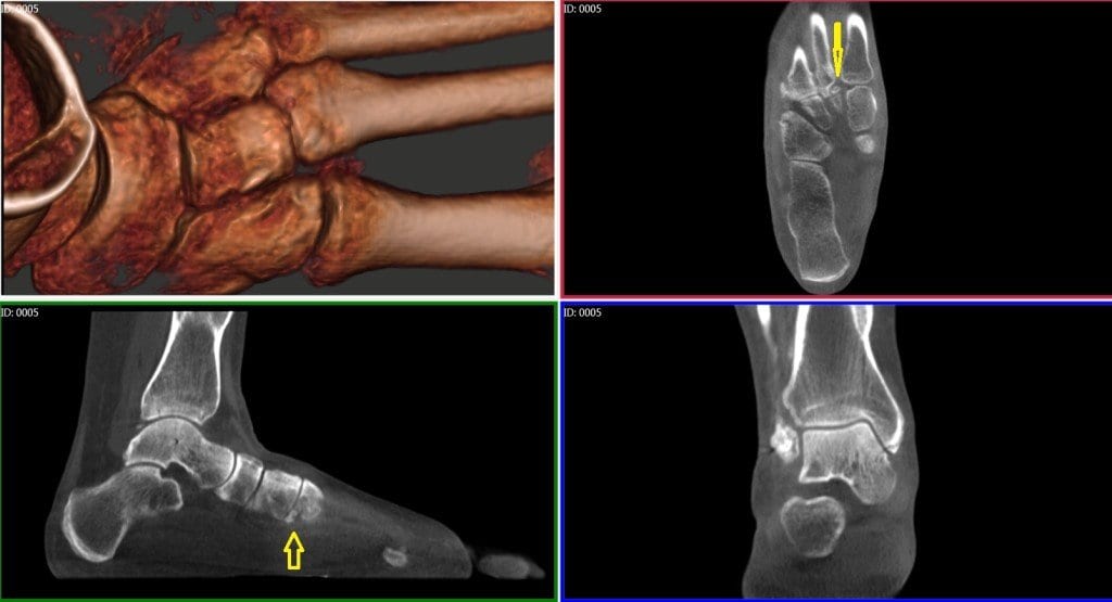
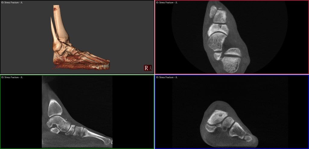
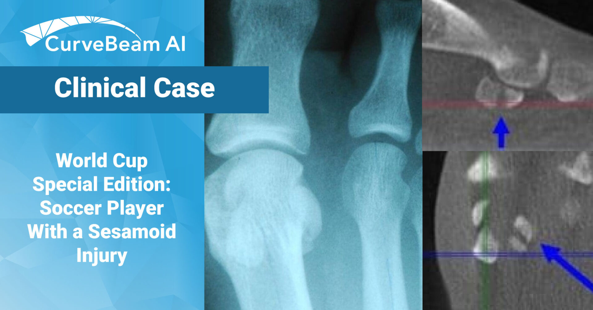
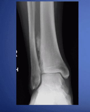
To see an example of a pedCAT scan that led to a more accurate diagnosis, click on the blog post title.

Read more

pedCAT: Easy to Incorporate into Your Medical Facility

The pedCAT system and your iPhone charger have something in common…you can plug them both in anywhere! That’s right. The pedCAT plugs into any standard 120 volt power outlet (or 220 volt outlet in certain countries). We suggest the pedCAT be connected to a dedicated line to prevent interference from other equipment as a precaution, although it is not required. The pedCAT’s simple power requirements are one of the factors that make it easy to integrate into any office workflow – even offices that have limited free space. Click on the blog post title to see some examples.
Read more

pedCAT: What’s the Dose?

At CurveBeam, patient safety is the top priority. When it comes to CT imaging, it’s important to know the radiation dose a patient can expect during the procedure.

So what is the radiation dose of a typical weight bearing CT scan taken with the pedCAT? But before we get to that, here’s a little quiz.

Can you rank the below items from greatest to least in terms of radiation exposure?

– A pedCAT scan of both feet

– A serving of Brazil nuts

– A roundtrip flight from New York to Tokyo

– An X-Ray of the chest

– Working in an Australian Uranium mine for a year

(Click the blog post title to see the answer.)

Read more
Back To Top