Orthopedic decision-making depends on accurate representation of anatomy—particularly when joint alignment and bone relationships change…

Weight-Bearing CT May Improve Diagnoses in Hallux Valgus Patients
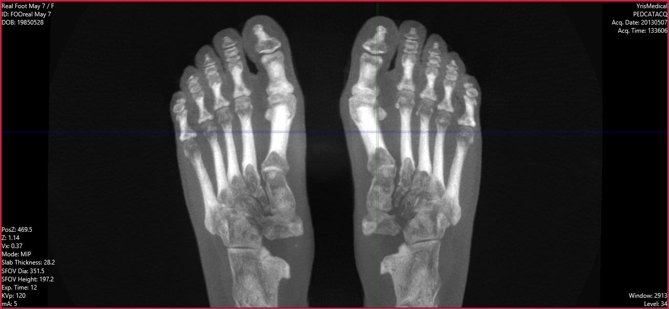
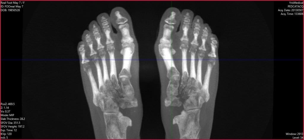
A recent analysis of studies titled Imaging of Hallux Valgus by James Welck and Naji Al-Khudairi has shown that newer, three-dimensional imaging techniques including weight-bearing computer tomography (WBCT) may permit a more thorough understanding of the hallux valgus (HV) deformity and provide greater information to foot and ankle surgeons prior to corrective surgery.
Traditional two-dimensional AP, lateral, and oblique radiographs are the current standard for imaging HV deformity. Two-dimensional radiographic images can be used to measure several radiographic angles which are commonly used to quantify the extent of HV deformity. However, recent research shows that some of these angle measures show low levels of reliability and accuracy. Coughlin et. al. found that measures of the hallux valgus angle and the intermetatarsal angle assessed using 2-D imaging had high intraobserver and interobserver reliability. In contrast, the distal metatarsal articular angle was measured to 5° or less in only 58.9% of cases, and measurement of metatarsophalangeal joint congruency led to a wide variation in cases identified. The primary reason that two-dimensional imaging techniques may fail to be reliable is that HV is a triplanar deformity with rotational aspects that two-dimensional images cannot account for.
Fortunately, research shows three-dimensional HV imaging techniques have promise. Conventional CT imaging is not weight-bearing, but weight-bearing cone beam CT provides better insight into HV. This means that many patients imaged through traditional CT may not have been diagnosed correctly.
- Welck and colleagues have found WBCT to better display the sesamoids, which are difficult to represent accurately using traditional AP radiographs.
- Collan and colleagues demonstrated the importance that CT is weight-bearing by finding that there were significant differences in HVAs and IMAs between HV and control groups only when patients were in a weight-bearing stance.
Two other research groups found that WBCT provided insight into the effects of joint hypermobility in HV patients which would not typically be measurable using two-dimensional imaging techniques.
To learn more about how three-dimensional imaging techniques like WBCT can benefit your foot and ankle practice, visit https://curvebeamai.com/products/pedcat/. CurveBeam’s PedCAT systems are at the cutting edge of WBCT innovation and can help provide your patients with the best HV care that current science has to offer.


