Orthopedic decision-making depends on accurate representation of anatomy—particularly when joint alignment and bone relationships change…
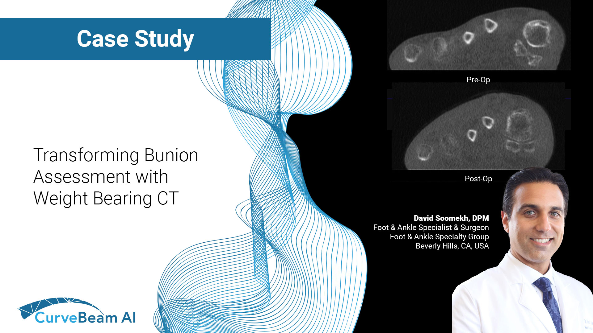
Transforming Bunion Assessment with Weight Bearing CT
By Dr. David Soomekh, DPM
Foot and ankle specialist and surgeon, sports medicine & reconstructive foot and ankle surgery, Foot & Ankle Specialty Group, Beverly Hills, CA
A Bunion (hallux abducto valgus) is a deformity involving the big toe and the bones associated with it. It is a deformity in 3 different planes. A bunion is the shift of the big toe bones into improper positions leading to pain and loss of function. The players involved in a bunion are: the big toe (hallux), the big toe joint (1st metatarsophalangeal joint), the 1st long bone (1st metatarsal), the sesamoids, the mid-foot joint (1st metatarsal-cuneiform joint), and the 1st mid-foot bone (1st cuneiform).
Over time, the 1st metatarsal will swing away from the other long bones towards the other foot (medial); this is what is seen as the bump on the side of the foot. At the same time, the big toe will move out of its joint towards the 2nd toe (lateral). Now the head of the 1st metatarsal bone is sticking out leading to undue pressure from shoes and the ground. This constant pressure and friction on the bone will cause extra bone to form, leading to the bump that is seen on the side of the foot getting larger. The big toe will continue to shift towards the 2nd toe causing an unbalanced big toe joint. This can cause arthritis to develop in the joint due to the mal-positioned joint. A bunion deformity is always progressive. It will always get worse over time. The sesamoids will also be in a poor position taking on improper pressure and causing sesamoiditis.
It is important when evaluating and treating a bunion deformity to understand that it is a 3 dimensional deformity. The clinical examination will help to understand the severity of the bunion and the areas of the patients pain due to the deformity. The first metatarsal phalangeal joint is examined for its range of motion and tracking. The first metatarsal cuneiform joint is evaluated for the degree of motion and how loose or hypermobile the joint is, or if there is any increased elevation of the first ray compared to the lessers rays.
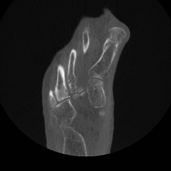
WBCT of AP view of bunion
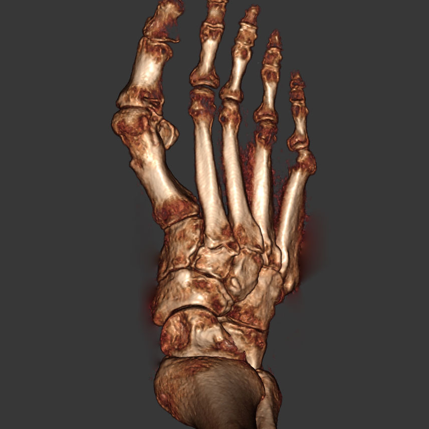
WBCT 3D view pre surgery
When surgically correcting a bunion deformity, it must be corrected in all three planes. Only correcting the transverse plane deformity will lead to failure of the correction and reoccurrence, especially with larger and hypermobile deformities. The first inter-metatarsal angle must be reduced. Any dorsal or plantar grade metatarsal a must be corrected. Any internal rotation of the first metatarsal must be de-rotated to allow for proper position of the sesamoids under the first metatarsal.
Radiographic examination is crucial to obtain a full picture of the bunion deformity. It is used to obtain the first inter-metatarsal angle and Hallux abducts angle in the AP view. With a lateral view one can elevate to some degree the amount of any elevatus of the first ray. The sesamoid view can give you minimal detail on the position of the sesamoids to evaluate rotation of the first metatarsal. Traditional radiographs, however, are limited in their ability to fully evaluate the three dimensional deformity.
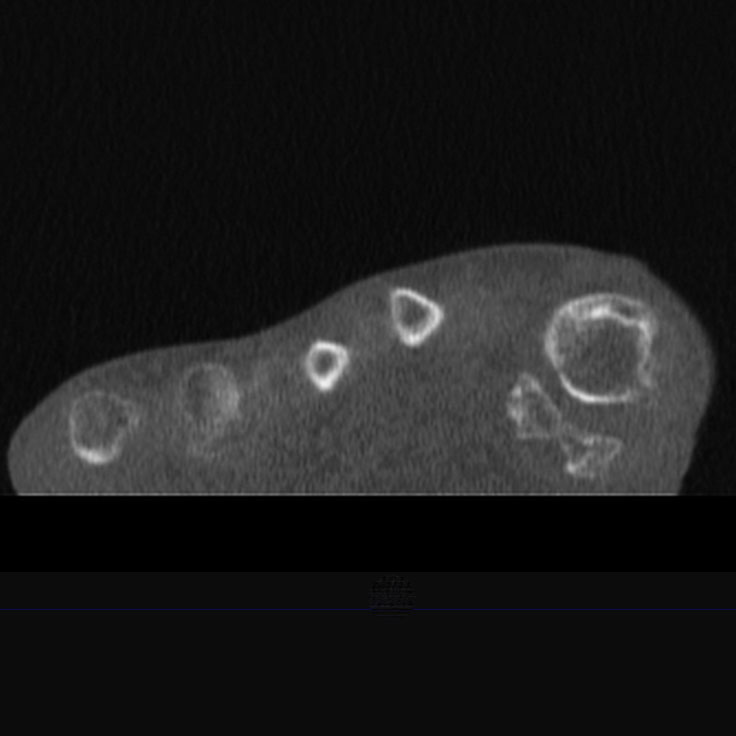
sesamoid view pre surgery
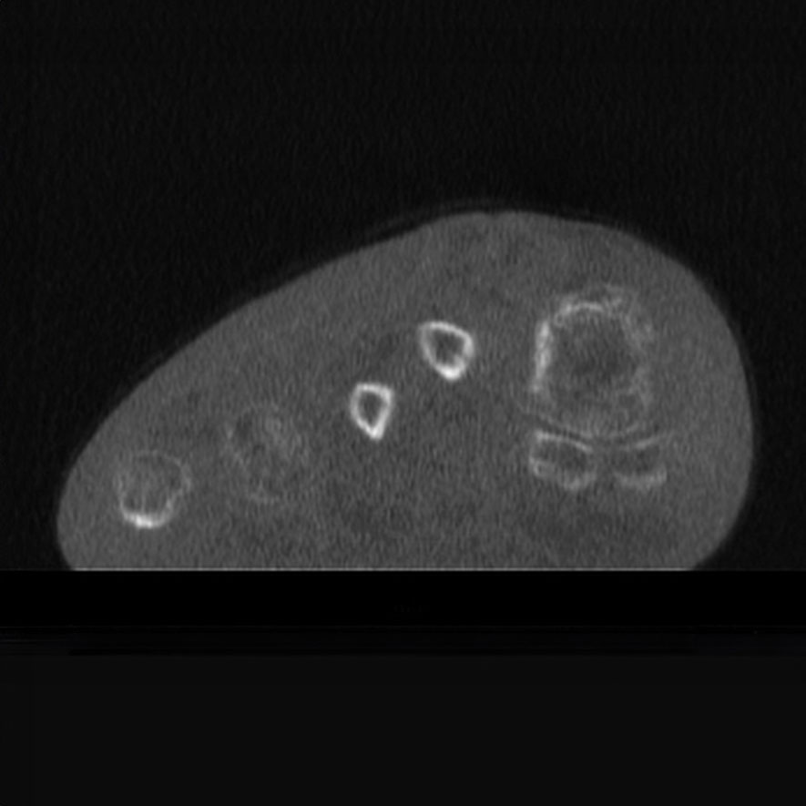
sesamoid view post surgery
A three dimensional image of the foot while weight-bearing is the ideal method to fully and precisely evaluate a bunion deformity for surgical planning. A weight bearing CT scan will give an exacting view in the X, Y, and Z planes in order to take measurements in all three planes. The most important view is the coronal view showing the position of the sesamoids under the first metatarsal and the degree of internal rotation of the first metatarsal.
In my practice when considering surgical correction of a Hallux Valgus deformity a weight bearing CT scan is always obtained. It is the most important tool that I use to precisely evaluate the deformity to plan my approach. It allows for a tailored surgical correction. Furthermore, I will use this CT scan post-operatively to confirm the proper amount of correction as well as the degree of bone healing.
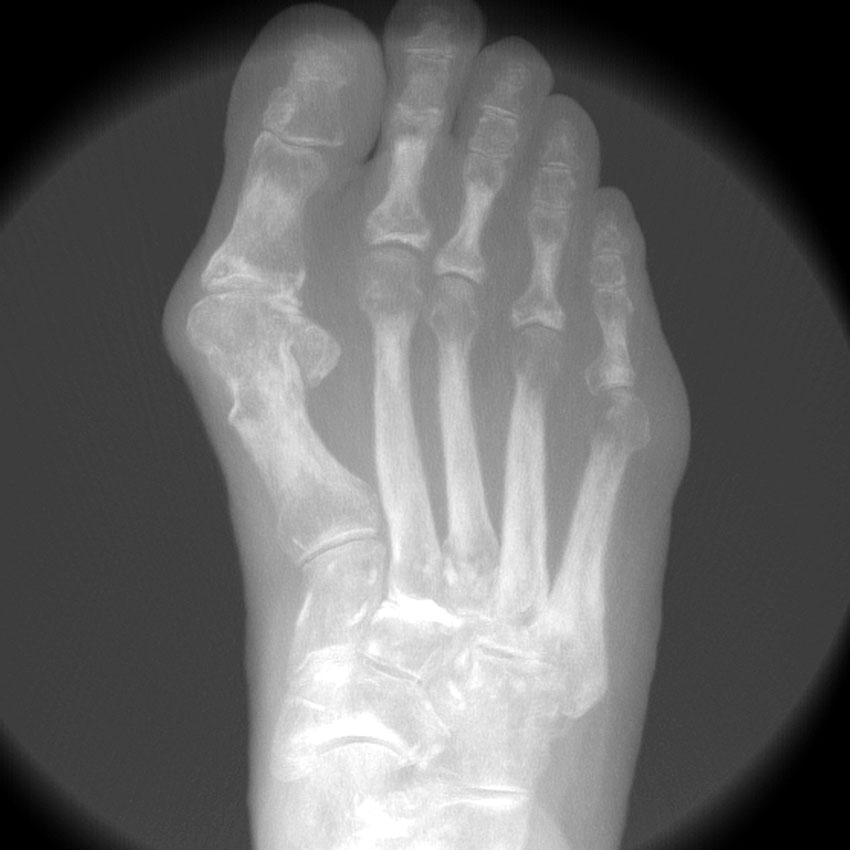
AP X Ray of bunion
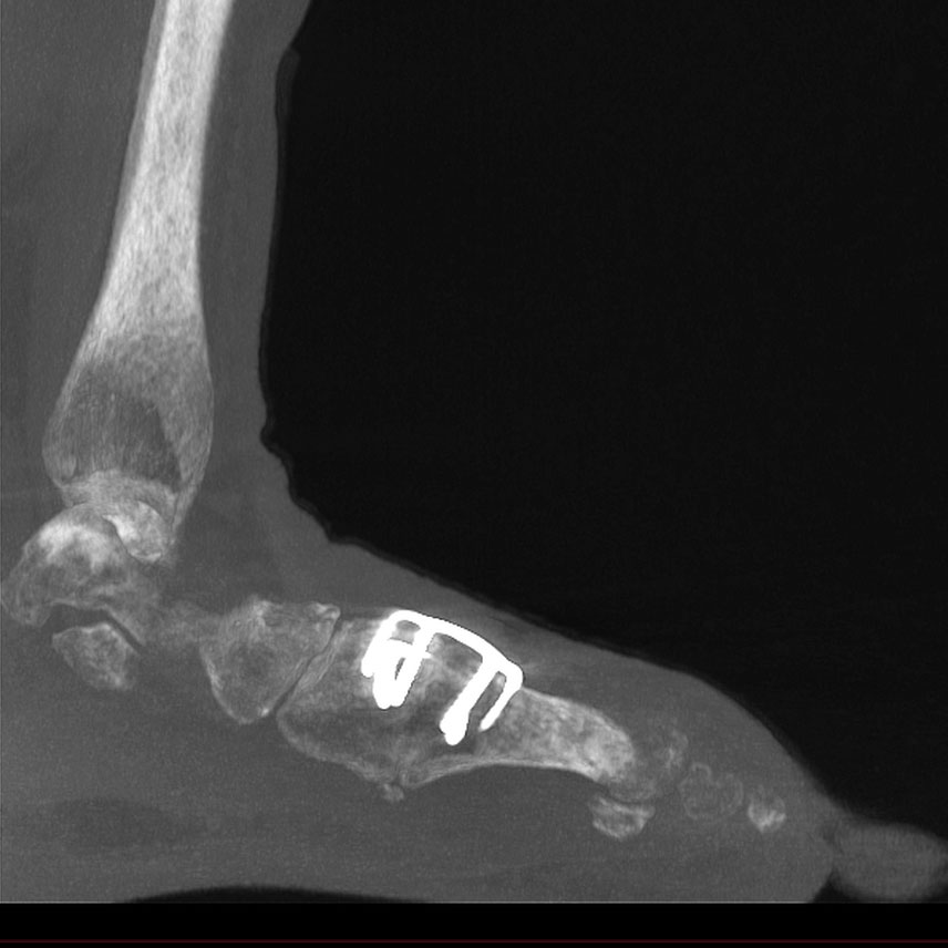
Post-surgical WBCT of Fused Lapidus
It has become especially useful when evaluating the fusion when I perform a Lapidus bunionectomy. I can be more confident that there is enough healing to allow for my patient to begin weight-bearing as well as when to be able to transition them into regular shoes. The use of the pedCAT scanner has improved my outcomes and decreased my complication rate.
CurveBeam AI’s Weight bearing CT is an essential tool for the highly trained and best foot and ankle surgeons for the evaluation and correction of hallux valgus.
Click here to listen to a podcast in which Dr. Soomekh discusses how CT became his primary in-office modality.


