AI-Driven Segmentation on Weight Bearing CT Introduction Accurate segmentation is critical for orthopedic workflows, including…
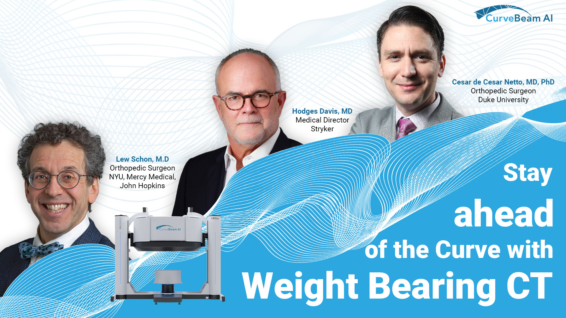
Stay ahead of the Curve with Weight Bearing CT
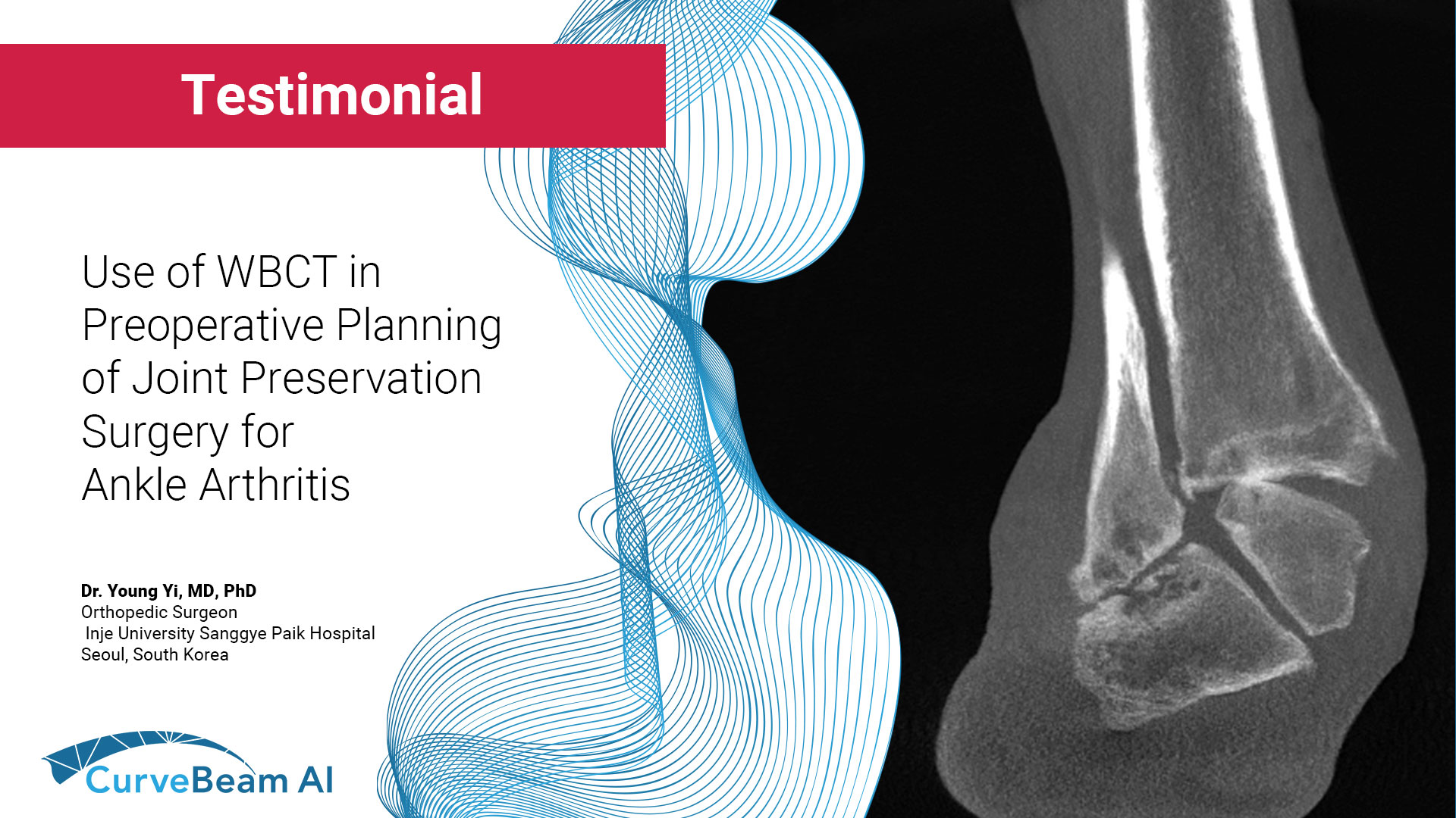
In the world of orthopedic diagnostics, imaging plays a crucial role in identifying deformities and planning surgical interventions. Conventional radiographs and MRIs have been the standard, but they come with limitations when it comes to understanding the complex, three-dimensional structure of the human body. This is where weight bearing CT (WBCT) emerges as a game changer.
Why Choose WBCT?
As highlighted in the video, traditional X-rays provide only a two-dimensional view of three-dimensional structures. Even weight bearing radiographs fail to capture the full relationships between bones. MRIs do not offer the same level of structural clarity in bone alignment under weight bearing conditions.
WBCT addresses these limitations by allowing clinicians to see the true alignment of bones in three dimensions—under the natural load of the patient’s body. This enables a far more accurate assessment of deformities, joint alignment, and overall foot and ankle biomechanics.
Enhancing Patient Care
For patients, the primary concern is simple: Why am I in pain, and how can I find relief? WBCT provides clearer answers by offering a comprehensive view of foot and ankle structures under load bearing conditions. This technology allows physicians to diagnose conditions with greater confidence and offer solutions that are more precise and effective.
A New Standard in Orthopedic Imaging
WBCT is transforming the way clinicians approach foot and ankle diagnostics. By providing unparalleled insight into bone alignment and structural relationships, this advanced imaging modality is setting a new standard for patient care. As more research continues to validate its benefits, WBCT is poised to become an essential tool in orthopedic and podiatric practices worldwide.
Stay ahead of the curve—embrace the future of imaging with WBCT.
Related Posts