Orthopedic decision-making depends on accurate representation of anatomy—particularly when joint alignment and bone relationships change…
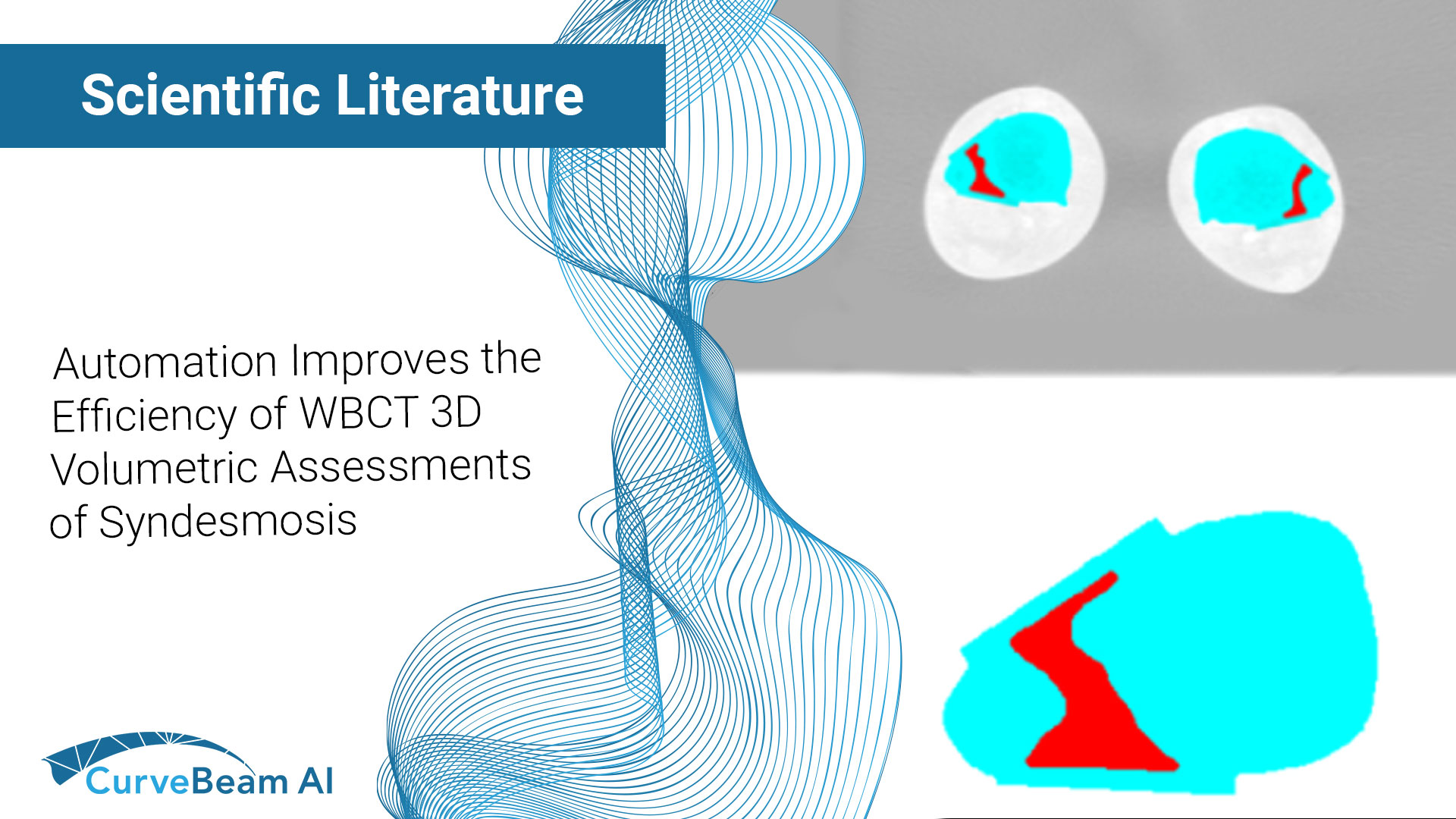
Automation Improves the Efficiency of Weight Bearing CT Scan 3D Volumetric Assessments of the Syndesmosis
Key Points:
- WBCT 3D Measurements volumetric measurements of the syndesmosis have shown to have a high specificity (83.3%) and sensitivity (95.8%) for the diagnosis of subtle injuries.
- Manual 3D WBCT volume measurement of syndesmosis, a promising method to detect instability, is tedious and does not have excellent interobserver agreement.
- Researchers at Massachusetts General Hospital have developed an automated volumetric measurement of the syndesmosis for weight bearing CT scans which takes only about 3 seconds and is more accurate and more reliable than manual methods to help clinicians in the diagnosis of ankle syndesmotic instability.
Incorporating these algorithms in the medical imaging software enables clinicians to run the measurements in a matter of seconds with a few clicks which make them a more convenient option given the crowded nature of the orthoapedic clinics.
Syndesmotic injuries are some of the most common foot and ankle injuries, particularly among athletes. Various diagnostic methods have been introduced for the detection of syndesmotic instability, in particular subtle syndesmotic ligamentous injuries leading to ankle instabilities.
Diagnosing Syndesmotic Injuries
X-Rays have typically been the first form of diagnosis; however, they have shown to not be completely reliable. CT scans outperform X-Rays, mainly for the detection of malreduction and for measuring diastasis. MRI is another option that while it has an almost perfect sensitivity and specificity of detection, it fails to assess subtle injuries due to its static nature. Ultrasound has shown promise as a cost-effective technique, but it has yet to reach its full diagnostic potential. As such, direct intraoperative assessment or arthroscopic examination has remained the gold standard for the diagnosis of instability, even though it is invasive and more costly.
WBCT has emerged as an effective diagnostic tool for the detection of syndesmotic instability. These promising results stem from the technology’s bilateral images of the joint under physiologic load while accounting for the 3D nature of this pathology. While this method does result in high specificity (83.3%) and sensitivity (95.8%) for the diagnosis of subtle instabilities, selecting the required anatomical landmarks is subjective and time consuming.
Dr. Soheil Ashkani-Esfahani et al out of the Foot & Ankle Research and Innovation Lab (FARIL), Department of Orthopaedic Surgery, Massachusetts General Hospital, Harvard Medical School, Boston, MA, USA hypothesized that automatized measurements would be more accurate and time-efficient than manual ones when applied to WBCT 3D scans.
Automated Algorithms for 3D WBCT Measurements
Researchers studied thirty cases of intraoperatively confirmed syndesmotic instability along with thirty individuals with no injuries to the ankle joint as cases and controls, retrospectively. Two observers conducted the manual volumetric measurements two times, at a one-week interval. An automated algorithm for 3D WBCT measurements was then developed to conduct the measurements on the axial images. Mann-Whitney U test was used to compared the values between human raters and computers then inter- and intra-class reliability was calculated.
Results showed a significant difference between the 3D syndesmotic volumes in cases and controls (12.6 ± 1.2 vs. 8.5 ± 1.9, respectively, p < 0.001). The ICC rate for the human observers was 0.75, while the automated method produced an ICC of 0.97. The Cronbach’s alpha test resulted in 0.60 and 0.62 in human observers, while the automated method produced 0.88. The mean time among human observers for 3D volume measurement of the syndesmosis in each patient was 268.4 ± 56.4 s (∼4–5 min) versus 2.9 ± 0.6 s by the automated measurement method (p < 0.001).
Conclusions
Researchers concluded that manual measurements are time-consuming and less precise than automatized methods. Automation of the 3D WBCT measurements seemed to be a reasonable step to a more accurate, time-efficient, and reliable method to help clinicians in the diagnosis of ankle syndesmotic instability.
It was noted that while the results were promising, 3D volumetric measurements are complex processes and need specific software. Clinicians will require simple methods. Researchers also stated the following:
- The raters in this study were experts with robust experience with 3D volume measurements of syndesmosis and that the timing of measurements may be different in providers with less experience.
- Their algorithm was designed on WBCT images and might not be useful in conventional CT scan images and unilateral images.
- They could not provide their algorithm online with free access to providers outside of their institution and that they would need to define rules and agreements to provide the algorithms online and available for all and deployable on the current image viewing software.
To read the full study click here.
Related Posts