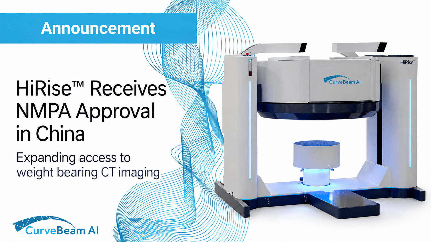
Expanding access to weight bearing CT imaging CurveBeam AI’s HiRise™ weight bearing CT system has…
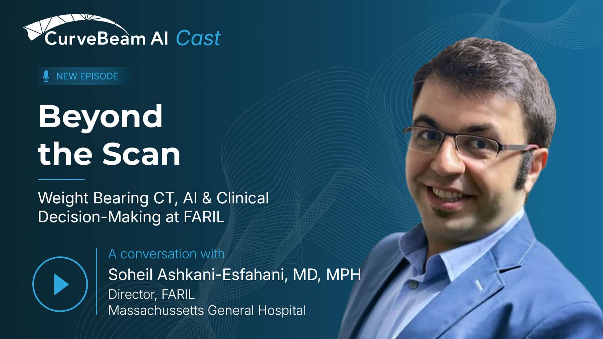
Beyond the Scan: How FARIL Uses Weight-Bearing CT to Support Research and Clinical Decision-Making
In the latest episode of CurveBeam AI Cast, Soheil Ashkani-Esfahani, MD, MPH, shares how FARIL connects clinical questions with advanced imaging, biomechanics, AI and engineering.
At a glance: At the Foot and Ankle Research and Innovation Laboratory (FARIL), weight bearing CT is more than an imaging exam. It provides quantitative, bilateral 3D data under physiologic load that can support the study of deformity and instability, patient-specific analysis, biomechanics, automated measurements, AI research, surgical simulation and clinical decision-making.
In the latest episode of CurveBeam AI Cast, host Vinti Singh speaks with Dr. Ashkani-Esfahani, Assistant Professor of Orthopedic Surgery at Harvard Medical School and Director of FARIL, about how the laboratory brings together clinical expertise, advanced imaging, biomechanics, engineering, and data science.
Their conversation looks beyond the scan itself. At FARIL, imaging can become the starting point for a larger process, one that begins with a clinical question and may lead to 3D analysis, biomechanical modeling, automated measurements, surgical simulation, patient-specific solutions or clinical study.
How FARIL Connects Clinical Problems With Research and Innovation
FARIL is a multidisciplinary laboratory designed to examine foot and ankle problems from multiple perspectives.
The team brings together orthopedic surgeons, research fellows, engineers, data scientists, radiologists, statisticians, and collaborators from around the world. Instead of treating imaging, biomechanics, AI, clinical outcomes, and device development as isolated areas, the lab seeks ways to connect them.
A project may begin with an imaging question, move into biomechanical or finite element analysis, inform the development of an automated measurement or algorithm, and ultimately support a clinical study or patient-specific solution.
FARIL also has an educational mission, providing research training and mentorship to medical students, residents, fellows, engineers, researchers, and visiting surgeons.
Across these initiatives, the goal remains consistent: to develop more accurate, personalized, and efficient approaches to caring for patients with foot and ankle disorders.
What Weight-Bearing CT Shows Under Natural Load
Weight bearing CT combines 3D imaging with physiologic loading, allowing clinicians and researchers to evaluate bones and joints while the patient is standing.
Dr. Ashkani-Esfahani refers to this as “natural loading” because it reflects how the patient’s anatomy behaves under the weight the patient can tolerate.
Traditional standing radiographs show the patient under load, but they compress complex anatomy into a two-dimensional projection. Conventional CT provides detailed 3D imaging, but it is generally acquired with the patient lying down, and the joints are not under physiologic load.
Weight bearing CT brings those two capabilities together. It provides a 3D view of bone and joint relationships while the patient is standing in a functional position.
This can help clinicians and researchers evaluate deformity, alignment, instability, joint-space relationships, bone position, and joint mechanics under load, relationships that may be difficult to appreciate fully on radiographs or non-weight bearing CT.
How FARIL Uses Weight-Bearing CT as Quantitative 3D Data
At FARIL, weight bearing CT is one of the central platforms connecting multiple research programs.
The laboratory uses the datasets to study conditions such as deformity, subtle instability, asymmetric joint wear, and changes in alignment. Because the imaging can be acquired bilaterally, the contralateral side may sometimes provide a patient-specific reference rather than requiring the team to rely entirely on population averages or standard cutoff values.
That comparison is not assumed to be valid in every case. FARIL is also studying normal side-to-side variability to better understand when the opposite limb can serve as a useful reference and when natural differences between sides need to be considered.
The 3D datasets can also support volumetric measurements and more detailed analysis of joint relationships. In the foot and ankle, Dr. Ashkani-Esfahani discusses applications involving conditions such as syndesmotic instability and Lisfranc instability, where subtle changes in alignment or joint position may be difficult to characterize using traditional methods alone.
“Weight bearing CT is not simply a diagnostic imaging tool. It is a quantitative 3D dataset that can support an entire research and clinical decision-making pipeline,” says Dr. Ashkani-Esfahani.
From a Surgeon’s Question to Patient-Specific Analysis
FARIL’s process begins with the clinical question, not with the technology.
A surgeon may bring the team a case involving a complex deformity, suspected instability, asymmetric joint wear, a previous reconstruction, a malunion, or an unclear relationship between the patient’s symptoms and the findings on conventional imaging.
The FARIL team can then systematically review the weight-bearing CT. That may include examining multiplanar images, comparing the involved and contralateral sides when appropriate, assessing 3D alignment, and evaluating joint relationships that may not be obvious on radiographs or conventional CT.
For selected cases, the analysis may go further.
The team can segment individual bones, create patient-specific 3D models, quantify deformity, or simulate a proposed correction. Researchers may also evaluate how osteotomy or reconstruction could affect alignment and joint loading.
The findings are then shared with the surgeon, who brings the clinical context, physical examination, patient symptoms, and operative considerations into the final decision.
This multidisciplinary approach does not replace clinical judgment. It provides the surgeon with additional patient-specific information to consider alongside the full clinical picture.
Extending the Dataset Through Biomechanics, AI and Surgical Planning
The weight bearing CT scan can also become the starting point for a broader research and analysis pipeline.
Patient-specific 3D bone models can be incorporated into:
- Finite element analysis
- Joint-contact and biomechanical studies
- Automated measurements
- Automated segmentation
- Machine-learning and deep-learning research
- Surgical simulation
- Patient-specific instruments and orthoses
One area Dr. Ashkani-Esfahani highlights is measurement reliability.
Even experienced clinicians may arrive at different values when performing measurements manually. Automated analysis has the potential to improve consistency by applying the same measurement method each time.
For researchers, this creates opportunities to study larger datasets more efficiently and to develop tools that may make complex 3D information easier to interpret and use.
For surgeons, patient-specific models and simulations may provide additional insight into the deformity, the intended correction, and how a proposed procedure could affect alignment or loading.
The episode illustrates how imaging, biomechanics, AI, and engineering can build on one another rather than functioning as separate technologies.
Diagnostic Confidence: Value a Traditional ROI May Miss
Not every scan will completely change the planned procedure. That does not mean it failed to add value.
Dr. Ashkani-Esfahani identifies diagnostic confidence as an important but less tangible benefit of advanced imaging.
A weight bearing CT scan may confirm that the surgeon has correctly understood the deformity and has considered rotational, contralateral or adjacent-joint factors that could otherwise be overlooked.
That confirmation can matter.
Clinical uncertainty may contribute to repeated radiographs, additional CT scans, delayed decisions, or disagreement among clinicians. Reaching a clear decision earlier may therefore create value even when the final treatment plan remains unchanged.
These benefits can be difficult to capture in a traditional ROI analysis that focuses primarily on procedure volume or imaging revenue. Dr. Ashkani-Esfahani’s perspective points toward a broader view of value, one that also considers the information available to the clinician, the efficiency of decision-making, and the confidence in that decision.
From Research Insight to Patient Care
At FARIL, the scan is not necessarily the endpoint.
It can become the foundation for multidisciplinary analysis, biomechanical research, patient-specific modeling, automated tools, clinical studies, and new approaches to surgical planning.
The full conversation offers a look at how one leading research laboratory is connecting these capabilities, and how quantitative 3D imaging under natural load can support a more complete understanding of the individual patient.
Watch the full CurveBeam AI Cast conversation with Dr. Soheil Ashkani-Esfahani.

Related Posts