At the 2025 American Alliance of Orthopedic Executives (AAOE) Annual Conference in Atlanta, CurveBeam AI…
How Weight Bearing CT Supports Orthopedic Collaboration in Hip, Knee, and Ankle Joint Replacement Cases
The take-away: Weight-bearing CT (WBCT) is increasingly being used in orthopedic surgical planning because it allows clinicians to evaluate lower extremity alignment in a functional standing position. For hip and knee arthroplasty surgeons, this additional 3D information may help support deformity assessment, preoperative planning, and cross-specialty collaboration with foot and ankle teams using a shared imaging dataset.
When patients present with pathology affecting multiple joints, surgical planning often extends beyond a single specialty. Hip, knee, foot, and ankle surgeons may all play a role in restoring alignment and function, making collaboration essential to achieving the best possible outcome.
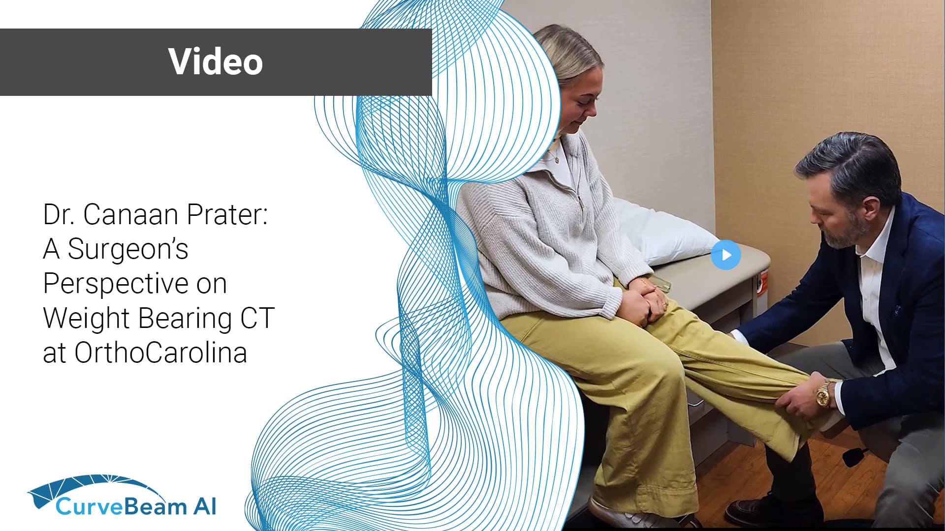
In a recent conversation, Dr. Canaan Prater, DO, hip and knee orthopedic surgeon at OrthoCarolina, discussed how weight bearing CT (WBCT) imaging supports a more comprehensive understanding of lower extremity alignment by capturing functional alignment under physiologic load.
“Weight bearing CT is a way to capture a three-dimensional image of the lower extremity in a way that the patient naturally is standing,” Dr. Prater explained. “It gets more of a physiologic positioning of alignment.”
Unlike conventional CT imaging performed in a non-weight-bearing position, WBCT allows orthopedic surgeons to evaluate how joints align and interact during functional standing conditions. For surgeons planning complex reconstructions, patient-specific approaches, or joint replacement procedures, this additional perspective can help support more informed decision-making.
A Shared View Across Orthopedic Specialties
Dr. Prater emphasized the value of using a single imaging study to support collaboration across orthopedic disciplines, particularly in patients who may require procedures involving the hip, knee, and ankle.
“There’ll be multiple cases where a patient comes in with pathology in both the hip and knee,” he said. “Many times they need both an ankle replacement and a knee replacement.”
By evaluating the same 3D weight bearing imaging dataset, surgeons across specialties can discuss alignment, sequencing, and surgical strategy using a shared anatomical reference point.
“This allows myself to collaborate with my foot and ankle colleagues,” Dr. Prater noted. “One image. We’re both looking at the same pictures.”
Supporting Surgical Planning
In addition to overall limb alignment, Dr. Prater described how WBCT imaging can help provide a more detailed understanding of tibial slope, femoral condyle deformities, and other alignment-related considerations relevant to hip and knee replacement planning.
“The key to the three-dimensional side of it gives me a much better and more accurate representation,” he said.
For orthopedic surgeons, access to additional anatomical information may help support individualized planning decisions while improving communication across care teams.
“It’s another tool that provides high-quality information,” Dr. Prater said. “The more information you have, the better decision you can make.”
Watch the full video interview with Dr. Canaan Prater below.
The views and opinions expressed in this video are those of Canaan Prater, DO, and do not necessarily reflect the official policy or position of CurveBeam AI. Any clinical insights or recommendations are based on his personal experience and judgment.
Related Posts