Fragility fractures are often the first visible sign of underlying osteoporosis but too often, they…
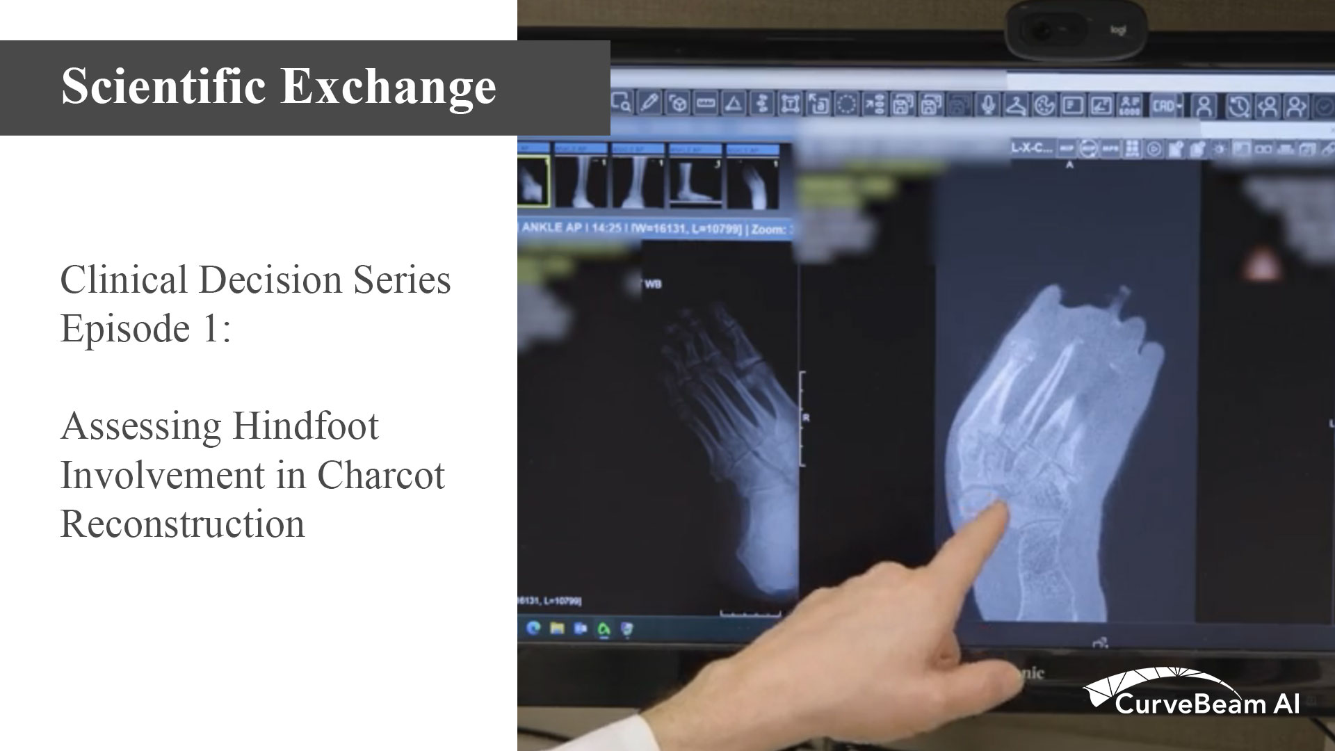
Clinical Decision Series, Episode 1: Assessing Hindfoot Involvement in Charcot Reconstruction
HiRise is cleared for 3D imaging of the upper and lower extremities and pelvis in adult and pediatric patients weighing 40 to 450 pounds. The device is intended to be operated in a professional healthcare environment by qualified healthcare professionals only. [View the FDA 510(k) summary and cleared indications for use.]
This material is provided for scientific exchange and is not a recommendation to use HiRise outside its cleared indications.
The Clinical Decision Series explores how physicians evaluate complex clinical questions by reviewing the examination findings, imaging information and treatment considerations involved in individual patient cases. Each episode focuses on the reasoning behind a specific clinical decision rather than prescribing a single approach.
In Episode 1 of the Clinical Decision Series, foot and ankle surgeon Lew Schon, MD, reviews the imaging and treatment of a 70-year-old patient with Charcot neuroarthropathy, severe midfoot collapse, foot abduction and a rocker-bottom deformity.
Dr. Schon compares conventional X-ray and weight-bearing CT (WBCT) images and explains how he interpreted the extent of midfoot and hindfoot involvement when developing the reconstruction plan.
This is a single-patient case presented to illustrate one physician’s clinical reasoning. It does not establish the safety or effectiveness of WBCT for Charcot reconstruction planning or demonstrate that the imaging produced the reported surgical or functional outcome.
Financial and Content Disclosure: Lew Schon, MD, is a paid consultant to CurveBeam AI. The clinical observations and opinions presented reflect Dr. Schon’s experience with this individual patient. This article and accompanying video were developed and edited by CurveBeam AI.
CASE OVERVIEW
Patient: A 70-year-old man with Charcot neuroarthropathy, midfoot collapse, foot abduction and a rocker-bottom deformity
Clinical Question: Had the Charcot-related joint destruction extended into the hindfoot, or was the hindfoot simply malaligned as a result of the severe midfoot collapse?
Radiographic Findings: Conventional weight-bearing radiographs demonstrated a severe deformity, but the complexity of the collapse made it difficult to determine the condition of the hindfoot joints.
WBCT Interpretation: Dr. Schon interpreted the WBCT images as showing extensive destruction localized to the midfoot while the hindfoot joints, including the subtalar joint, remained preserved.
Surgical Strategy: Based on his overall clinical evaluation and imaging assessment, Dr. Schon elected to reconstruct the midfoot while preserving the hindfoot.
WHAT WAS DIFFICULT TO DETERMINE ON THE X-RAYS?
The patient’s radiographs demonstrated marked midfoot collapse with significant abduction and a classic rocker-bottom deformity. While the overall severity of the deformity was readily apparent, the overlapping anatomy made it challenging to determine whether the hindfoot joints themselves had been affected by the Charcot process.
As Dr. Schon explains in the video, the central question was whether the hindfoot required reconstruction because of true joint destruction or whether its appearance simply reflected secondary malalignment caused by the collapsed midfoot.
Answering that question would significantly influence the surgical approach.
WHAT DID DR. SCHON OBSERVE ON THE WEIGHT-BEARING CT IMAGES?
According to Dr. Schon, the WBCT images allowed him to evaluate the deformity in three dimensions and better distinguish the midfoot findings from the condition of the hindfoot joints. He interpreted the scan as demonstrating extensive collapse within the midfoot while showing preservation of the subtalar joint and the remaining hindfoot articulations despite their concerning appearance on conventional radiographs.
Dr. Schon also identified a localized area of impingement and a cyst. In his assessment, these represented focal findings rather than widespread destruction of the hindfoot joint complex.
HOW DID THE IMAGING FACTOR INTO THE RECONSTRUCTION PLAN?
After integrating the patient’s physical examination with the imaging findings, Dr. Schon planned the reconstruction around the midfoot while preserving the hindfoot joints.
“You need to see that this hindfoot is okay or not okay. So the weight-bearing scan really makes a difference here.”
Lew Schon, MD
The operative footage demonstrates reconstruction and stabilization of the midfoot while leaving the hindfoot intact. According to Dr. Schon, preserving those joints allowed the patient to maintain hindfoot motion and avoid the additional stiffness associated with a more extensive fusion.
WHAT POSTOPERATIVE FINDINGS DOES DR. SCHON REPORT?
Postoperative imaging demonstrated correction of the rocker-bottom deformity and substantial improvement in foot alignment. Dr. Schon also reports that the patient’s hindfoot and ankle remained intact throughout the follow-up period discussed in the video.
He further reports that the patient was able to walk, climb stairs and wear regular shoes without the need for an ankle-foot orthosis (AFO), walker or wheelchair.
These observations reflect the treating surgeon’s reported outcome for this case and were not collected as part of a controlled clinical study.
HOW DOES THIS CASE RELATE TO THE PUBLISHED LITERATURE?
This case highlights a clinical question that continues to be explored in the literature: how three-dimensional imaging under physiologic load may contribute to the evaluation of complex Charcot deformity.
A 2026 narrative review by Kim and Yi describes several potential applications of WBCT in Charcot neuro-osteoarthropathy, including three-dimensional assessment of osseous alignment during weight bearing and preoperative surgical planning. The authors also discuss important limitations, including imaging artifacts, reduced soft tissue visualization compared with MRI, variability in acquisition and measurement techniques, cost and limited availability. They conclude that prospective studies are needed to better understand how WBCT-derived measurements relate to long-term clinical outcomes. The review received no external funding, and the authors reported no conflicts of interest.[1]
A separate 2026 retrospective study by Bolisetti and colleagues compared conventional weight-bearing radiographic measurements with three-dimensional WBCT measurements in seven patients with Charcot neuroarthropathy. The investigators reported differences between several two-dimensional and three-dimensional measurements but did not evaluate surgical decision-making or patient outcomes.[2]
ADDITIONAL SCIENTIFIC INFORMATION ABOUT WBCT AND CHARCOT NEUROARTHROPATHY
Visit our Scientific Literature page here.
References
1. Kim NY, Yi Y. The Role of Weight-Bearing Computed Tomography in the Assessment and Management of Charcot Foot Deformity: A Narrative Review. *Medicina*. 2026;62(1):117. doi:10.3390/medicina62010117. [Access the free full-text article.]
2. Bolisetti A, Koroneos Z, Chopra AA, et al. Two-Dimensional vs Three-Dimensional Analysis of Weightbearing Charcot Neuropathy Deformities. *Journal of Foot and Ankle Surgery*. 2026;65(3):101.e1-101.e7. doi:10.1053/j.jfas.2026.01.010. [View the publication record and abstract.]
3. U.S. Food and Drug Administration. HiRise 510(k) Premarket Notification K241713. [View the FDA 510(k) summary and indications for use.]

Related Posts